
Protrusio acetabuli is medial migration of the femoral head, with the acetabular line crossing the ilioischial line on an AP pelvis radiograph. Causes include rheumatoid arthritis, Paget disease, osteomalacia and Marfan syndrome. Hip replacement needs medial bone grafting and restoration of the hip centre.
Definition
- Medialisation of the medial wall of the acetabulum with a centre-edge angle > 40° on an AP XR of the pelvis
- Result of remodelling of weak, medial acetabular bone after multiple, recurring stress fractures
- Most often resulting secondary to inflammatory cause (inflammatory destruction) or metabolic cause (qualitative deficiency in the bone)
- Migration occurs along the resultant joint-reaction force vector (McCullum et al 1980)
Aetiology
Primary
- Otto Pelvis (Arthrokatadysis)
- Bilateral 1/3rd
- Middle aged females
- Pain & Decreased ROM early
- Coxa Vara & Osteoarthritis common
- ? Causally related to osteomalacia
- Diagnosis of exclusion
Secondary
- Inflammatory arthritis
- 15% of Rh arthritis of the hip
- 33% of ankylosing spondylitis of the hip
- Metabolic
- Osteomalacia – up to 50%
- Renal osteodystrophy
- Hyperparathyroidism
- Paget’s disease
- Osteoporosis
- Connective Tissue disorder
- Marfan’s
- Erler-Danlos
- Osteogenesis imperfecta
- Traumatic
- Central fracture-dislocation
- Septic arthritis
- Neoplastic
- Existing Prosthesis
- Hemiarthroplasty
- Seen in 5% of those with Osteoarthritis
Clinical Findings
- Progressive pain & stiffness in groin
- Limp
- Trendelenburg +
- Antalgic gait
Investigations
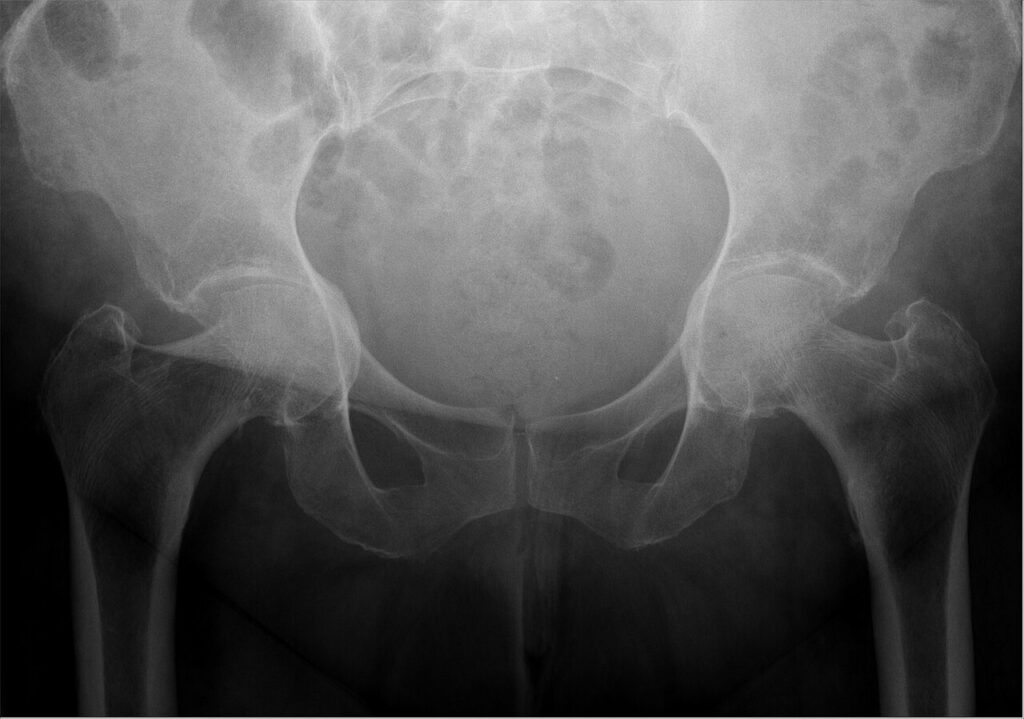
X-ray
- Position of femoral head in relation to medial wall acetabulum & pelvic brim = iliopectineal line
- Grade I Mild 1-5mm
- Grade II Moderate 6-15mm
- Grade III Severe > 15mm
- Medial wall (acetabular line) & it’s relation to ilioischial line (Kohler’s line)
- Men Normal = acetabular line 2mm lateral to I-I line
- Women Normal = 1mm medial to I-I line
- Grade I 3-8mm 6-11mm
- Grade II 8-13mm 12-17mm
- Grade III > 13mm > 17mm
- CE > 40°
Blood
- FBC
- ESR
- CRP
- RhF
- ANA
- Se electrolytes
Synovial Bx
- Indicated when inflammatory arthritis or infection is suspected but unconfirmed
- Histology identifies rheumatoid synovitis and tuberculous granulomas
- Send tissue for culture including mycobacterial culture
- Obtained open, arthroscopically or by image guided needle biopsy
Treatment
Skeletally Immature
- Triradiate epiphyseodesis
- If coxa varus present then consider adding valgus intertrochanteric osteotomy
Skeletally Mature Adolescent/ Young Adult
- Valgus intertrochanteric osteotomy
- Indications
- significant symptoms with minimal degenerative changes in patient < 40 years
- Principle
- redirect joint-reaction force superiorly
- Pointers
- Amount of correction determined by amount of preoperative adduction
- Generally 20-30° correction desirable
- Lateralisation of the femur to correct mechanical alignment
- Soft-tissue release (esp psoas) to effect lateralisation & improve hip ROM
- Arthroplasty
- Replacement
- Resection
- Interposition
- Arthrodesis
Older Adult
- THR
- THR in protrusio acetabuli
- Principles
- 1. Place hip centre in anatomic location
- To restore biomechanics
- 2. Restore bone stock
- Reconstruct segmental/ cavitatory defects of medial wall with bone graft
- Use intact peripheral rim for component fixation
- 1. Place hip centre in anatomic location
- Preoperative
- Define acetabular defect with appropriate imaging ~ 3D CT
- Template preoperative LLD
- Intraoperative
- Sciatic nerve should be identified as closer to joint
- Trochanteric Osteotomy may be required for exposure
- Dislocation may be very difficult
- Removal of rim bone – first try removing portion of posterior acetabulum
- Head delivery piecemeal
- May have to do neck osteotomy in situ
- Significant soft tissue release from femur needed
- Don’t ream medially or use centring hole
- Remove cartilage & soft tissue with curette
- Enlarge rim only with reamer
- Take care not to create peripheral defect
- Use one or more techniques to prevent medial migration
- Use wire mesh in floor
- Graft floor with morsellized bone
- Cup may be
- Uncemented rim fit cup
- Cemented cup into impaction graft
- Cemented cup into protrusio ring
- Large cup with flange
- Outcome
- adequacy of correction of the deformity correlates with long-term prosthetic survival
- Medial cup placement lead to high medial stresses cf. anatomic placement
- Metal-backed component effective due to superior stress dissipation of the metal cup
- Metal protrusio ring more reliably transfers stress from the medial wall to the rim than does protrusio cup
- Reinforcement of medial wall with cement & wire mesh not effective
- 50% reconstructed hips with cup centre > 10mm from anatomic hip centre failed
- Increased loosening cup with protrusio grade 1
- Multiple studies have shown excellent results with bone graft used to (1) lateralise the cup & (2) restore bone stock (usually femoral head morcellized or fragmented with or without cement)
- Most widely used approach today is fill defect with morcellized graft then use porous coated cup
- Ranawat & Zahn
- Principles
- THR in protrusio acetabuli
| Degree of Protrusi | Management |
|---|---|
| Protrusio < 5mm | THR without BG |
| Protrusio > 5mm with intact medial wall | THR with BG |
| Protrusio > 5mm with deficient medial wall | THR with BG & additional fixation (uncemented cup with screw fixation or antiprotrusio ring) |
Reviewed by Professor Phong Tran, Head of Orthopaedic Surgery, Western Health. Last updated 10 October 2026.