
Femoroacetabular impingement is abnormal contact between the proximal femur and the acetabular rim, causing labral and cartilage damage. Cam morphology is an aspherical head neck junction and pincer morphology is acetabular overcoverage. Symptomatic patients without arthritis may benefit from arthroscopic or open osteochondroplasty.
Overview
- Abnormal contact between proximal femur and acetabular rim, causing labral and chondral damage
- Cam is an aspherical head neck junction, pincer is acetabular overcoverage, many have both
- Young active patients with groin pain on flexion and internal rotation, positive FADIR
- AP pelvis and lateral views, MR arthrography for labrum and cartilage
- Physiotherapy first line
- Osteochondroplasty with labral repair for persisting symptoms without advanced arthritis
Western Health Orthopaedic Registrar presentation – FAI and Labral Pathology of the Hip by Dr Peter Moore
Classification
| Type | Description |
|---|---|
| Cam | Aspherical anterosuperior head neck junction, young male athletes, after SCFE or Perthes disease |
| Pincer | Global (coxa profunda, protrusio) or focal (retroversion) overcoverage, more common in women |
| Mixed | The most common pattern |
Original publication Ganz R, Parvizi J, Beck M, Leunig M, Nötzli H, Siebenrock KA. Femoroacetabular impingement: a cause for osteoarthritis of the hip. Clin Orthop Relat Res. 2003(417):112-20.
- Cam morphology is common in asymptomatic people
Pathology
- Cam causes outside in anterosuperior delamination (carpet lesion) and chondrolabral separation
- Pincer crushes the labrum, causing degeneration, ossification and posteroinferior contrecoup injury
Clinical Features
- Groin pain with C sign, worse with sitting, deep flexion and pivoting
- Reduced internal rotation in flexion
- FADIR is sensitive but not specific
Investigations
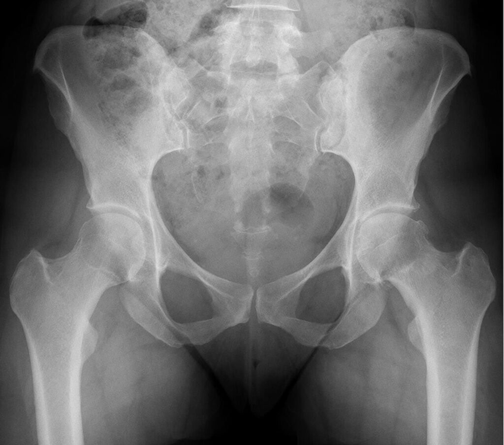
- Alpha angle above about 55 to 60 degrees and pistol grip deformity indicate cam
- Crossover, posterior wall and ischial spine signs indicate retroversion
- LCEA above about 40 degrees and negative Tönnis angle indicate overcoverage
Management
- Arthroscopic osteochondroplasty, rim trimming, labral repair and capsular repair
- Ganz surgical dislocation for complex or global deformity
- Anteverting PAO for retroversion with posterior wall deficiency
- UK FASHIoN found arthroscopy improved hip quality of life more than physiotherapy
- Poor results with Tönnis 2 or more, joint space 2 mm or less, dysplasia or extensive chondral damage
Complications
- Residual deformity, the commonest reason for revision
- Traction neurapraxia, heterotopic ossification and iatrogenic instability
Reviewed by Professor Phong Tran, Head of Orthopaedic Surgery, Western Health. Last updated 10 October 2026.