
Question
A 15 year old female gymnast has 6 months of low back pain made worse by extension. A lateral radiograph shows a forward slip of L5 on S1.
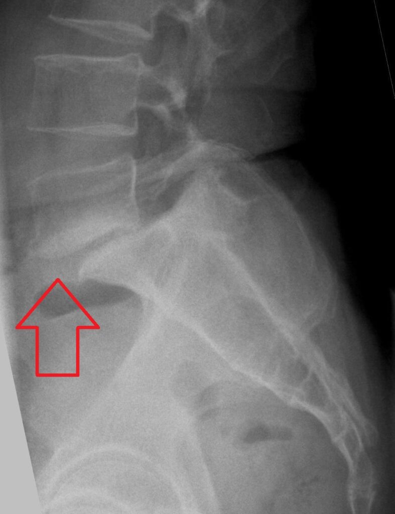
- What is the diagnosis and how is it classified?
- What radiographic parameters would you measure?
- How would you manage her?
Answer
Diagnosis and classification
- Isthmic spondylolisthesis from bilateral L5 pars interarticularis defects (spondylolysis)
- Stress fracture from repetitive hyperextension in gymnasts, fast bowlers and divers
- Wiltse classification types are dysplastic, isthmic, degenerative, traumatic, pathological and iatrogenic
- Meyerding grades I to IV by percentage slip (0 to 25, 25 to 50, 50 to 75, 75 to 100%), with grade V spondyloptosis
Radiographic parameters
- Slip percentage
- Slip angle, with values over 10 degrees predicting progression
- Pelvic incidence, which is higher in spondylolisthesis
- Lumbosacral kyphosis and a dome shaped sacrum or trapezoidal L5 in high grade slips
- Oblique radiographs show the collar on the Scottie dog, and CT or SPECT shows acute pars lesions
Management
- Low grade, activity modification, core strengthening and a brace for acute painful pars lesions
- Pars repair for symptomatic lysis without slip at L1 to L4
- In situ posterolateral fusion of L5 to S1 for persistent pain or progressive slip over 50%
- High grade slips may need L4 to S1 fusion and reduction, with risk of L5 nerve root injury
- Monitor growing patients for progression
Related pages
Author Contributions
Orthofracs team
Reviewed by Professor Phong Tran, Head of Orthopaedic Surgery, Western Health. Last updated 10 October 2026.