
Question
A 38 year old office worker has six weeks of left leg pain radiating to the lateral foot. He has weak plantarflexion and an absent ankle jerk.
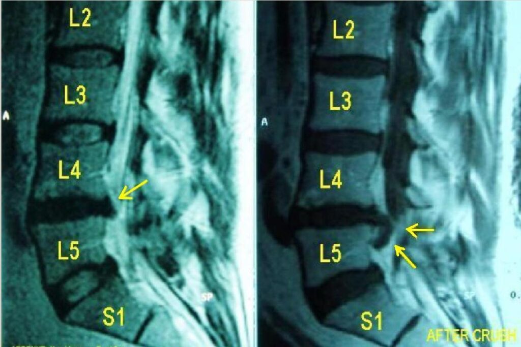
- Which root is affected and what does the MRI show?
- What red flags would change your management?
- Discuss non-operative care and the evidence for surgery.
Answer
Root and imaging
- S1 radiculopathy, weak gastrocnemius, absent ankle reflex, lateral foot numbness
- L5 to S1 disc prolapse with extrusion
- Posterolateral disc at L5 to S1 compresses the traversing S1 root
- Far lateral disc at L4 to L5 compresses the exiting L4 root
Red flags
- Cauda equina symptoms, saddle anaesthesia, bladder or bowel change
- Progressive or profound motor deficit
- Fever, weight loss, cancer history or immunosuppression
- Night pain unrelated to posture
Management
- Most improve over 6 to 12 weeks
- Analgesia, activity as tolerated, physiotherapy
- Transforaminal epidural steroid injection for short term relief
- Microdiscectomy for persistent pain beyond 6 to 12 weeks or progressive weakness
- SPORT showed faster relief with surgery, with groups converging over time
- Peul (2007) showed faster recovery with early surgery and similar results at one year
- Recurrent herniation after discectomy about 5 to 10%
Related pages
Author Contributions
Orthofracs team
Reviewed by Professor Phong Tran, Head of Orthopaedic Surgery, Western Health. Last updated 10 October 2026.