
Question
A 78 year old woman on long term corticosteroids has severe low back and buttock pain with no history of trauma. Plain radiographs are normal.
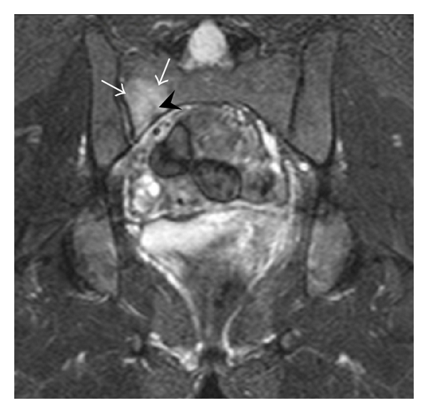
- What does the MRI show?
- What imaging signs are described?
- Outline management.
Answer
MRI
- Bone marrow oedema in the right sacral ala
- Sacral insufficiency fracture
- Risk factors include osteoporosis, corticosteroids, pelvic radiotherapy, rheumatoid arthritis and prior lumbar fusion
Imaging signs
- Radiographs often normal
- Honda sign (H pattern) on bone scan when bilateral with a horizontal limb
- CT shows sclerosis and fracture lines
- MRI is most sensitive
- Look for pubic rami fractures, which coexist in many
Management
- Analgesia and mobilisation as tolerated
- Investigate and treat osteoporosis, vitamin D and calcium
- Consider teriparatide, which may speed healing
- Sacroplasty for refractory pain
- Percutaneous iliosacral or transsacral screw fixation for displaced or non healing fractures
- Thromboprophylaxis while mobility is limited
Related pages
Author Contributions
Orthofracs team
Reviewed by Professor Phong Tran, Head of Orthopaedic Surgery, Western Health. Last updated 10 October 2026.