
Question
A 66 year old woman with longstanding rheumatoid arthritis has occipital headaches, clumsy hands and is falling more often.
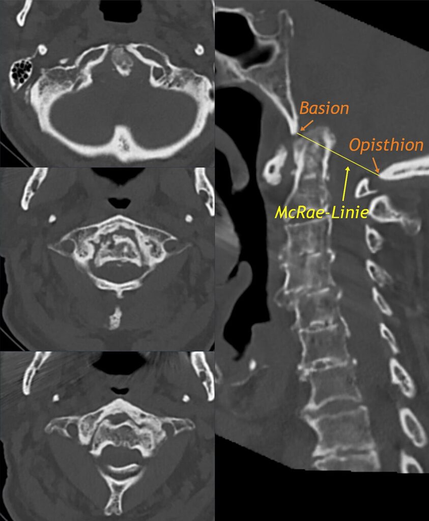
- What does the CT show?
- Describe the three patterns of rheumatoid cervical instability and how each is measured.
- What are the indications for surgery?
Answer
CT
- Odontoid tip projects above the foramen magnum
- Basilar invagination (cranial settling)
- Risk of brainstem and upper cord compression
Patterns of instability
| Pattern | Measurement |
|---|---|
| Atlantoaxial subluxation (most common) | ADI over 3.5 mm. PADI under 14 mm predicts paralysis |
| Basilar invagination | Ranawat under 13 mm, Redlund-Johnell under 34 mm (men) or 29 mm (women), McGregor line breached by over 4.5 mm |
| Subaxial subluxation | Stepladder subluxation, canal under 14 mm |
Surgery
- Myelopathy, as natural history is poor once established
- Intractable pain
- PADI under 14 mm or canal under 14 mm even without symptoms
- Cranial settling with brainstem compression
- C1 to C2 fusion for reducible atlantoaxial subluxation
- Occipitocervical fusion for basilar invagination, transoral odontoid resection if irreducible
- Flexion and extension views before any elective anaesthesia
Related pages
Author Contributions
Orthofracs team
Reviewed by Professor Phong Tran, Head of Orthopaedic Surgery, Western Health. Last updated 10 October 2026.