
Question
A 30 year old woman falls onto her outstretched hand during netball.
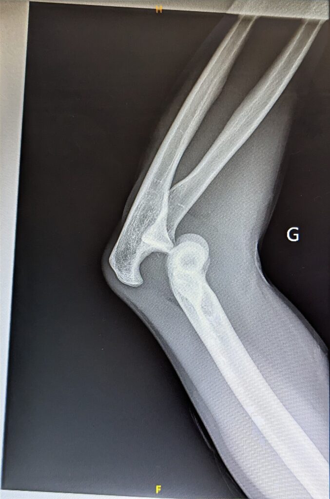
- Describe the radiograph and the mechanism.
- How would you reduce it and assess stability?
- Outline rehabilitation and the indications for surgery.
Answer
Radiograph and mechanism
- Posterolateral dislocation of the elbow without fracture (simple dislocation)
- Axial load, valgus, supination and external rotation
- Horii circle, injury progresses from the LCL complex anteriorly and posteriorly to the MCL
- LUCL injury is the primary lesion
Reduction and assessment
- Document median, ulnar and radial nerve function and pulses
- Sedation, correct medial or lateral shift, then traction with forearm supinated and elbow flexed
- Check range after reduction, note the extension at which it redislocates
- Post reduction films to confirm a concentric joint and exclude fractures
- CT if fractures of the coronoid or radial head are suspected
Rehabilitation
- Short period of splinting for comfort, under one week
- Early active motion within the stable arc, overhead motion protocol
- Pronation protects LCL deficient elbows
- Prolonged immobilisation over 3 weeks causes stiffness
Surgery
- Elbow unstable within 30 degrees of extension despite pronation
- Incarcerated fragments or non concentric reduction
- LCL repair to the lateral epicondyle, then MCL repair if still unstable
- Hinged external fixator for persistent instability
Related pages
Author Contributions
Orthofracs team
Reviewed by Professor Phong Tran, Head of Orthopaedic Surgery, Western Health. Last updated 10 October 2026.