
Question
A 50 year old builder has pain at the end of elbow extension and loss of the last 30 degrees of extension. He has intermittent locking.
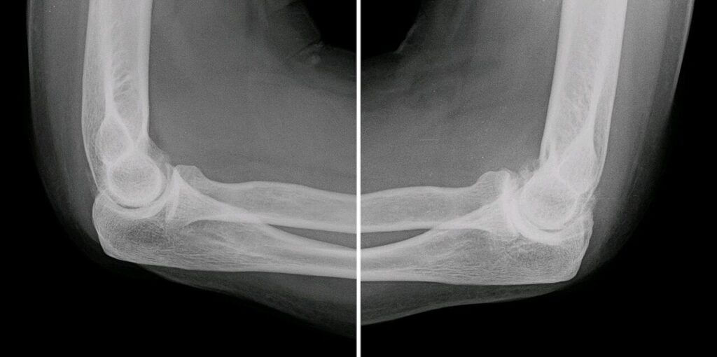
- Describe the radiograph and the typical presentation.
- What else would you assess?
- Outline the treatment options.
Answer
Radiograph and presentation
- Osteophytes at the olecranon tip, coronoid and their fossae
- Relative preservation of the ulnohumeral joint space
- Loose bodies
- Primary elbow osteoarthritis, a disease of heavy manual workers and weightlifters
- Pain at the end of range, mid arc is pain free
Further assessment
- Ulnar nerve symptoms, present in up to half
- CT with 3D reconstruction to map osteophytes and loose bodies
- Exclude inflammatory or post-traumatic arthritis
Treatment
- Activity change, analgesia and injection
- Arthroscopic debridement and loose body removal
- Open osteocapsular arthroplasty (Outerbridge-Kashiwagi or column procedure)
- Ulnar nerve decompression or transposition when symptomatic or flexion under 100 degrees before release
- Interposition arthroplasty in young patients with advanced disease
- Total elbow arthroplasty in older low demand patients only, lifting limit about 2 to 5 kg
Related pages
Author Contributions
Orthofracs team
Reviewed by Professor Phong Tran, Head of Orthopaedic Surgery, Western Health. Last updated 10 October 2026.