
Complex elbow instability is dislocation with associated fractures, including the terrible triad of dislocation with radial head and coronoid fractures, and varus posteromedial rotatory instability. Surgery restores the coronoid, radial head and lateral ligament complex so the elbow stays stable through early motion.
Video
Definition
- Condition resulting from injury & resultant loss of function due to damage to the articular surface & ligamentous structures that stabilise the elbow
- May present as subluxation or incongruity with malalignment
Aetiology
- Fall onto an outstretched hand with axial load, supination and valgus
- High energy trauma such as falls from height and road trauma
- Varus posteromedial rotatory load producing anteromedial coronoid facet fracture
- Iatrogenic LCL injury during lateral approaches or after radial head excision
Epidemiology
- Elbow is the second commonest dislocated major joint in adults after the shoulder
- Elbow dislocation incidence about 5 per 100,000 per year
- Peak in young adults from sport, second peak in older adults from falls
- Simple dislocations are more common than complex fracture dislocations
Anatomy
- Ulnohumeral, radiocapitellar and proximal radioulnar joints share one capsule
- Coronoid resists posterior translation, anteromedial facet resists varus
- Radial head is a secondary restraint to valgus and posterolateral rotation
- LUCL inserts on the supinator crest of the ulna
- Anterior bundle of the MCL inserts on the sublime tubercle
Contributions to Normal Stability
Static Constraints
- Primary
- Ulnohumeral articulation
- Medial collateral ligament
- Lateral collateral ligament
- Secondary
- Radial head
- Common flexor tendon
- Common extensor tendon
- Capsule
Dynamic Constraints
- Muscles
- that cross the elbow – providing compression
- Especially
- Triceps
- Anconeus
- Brachialis
- articular surfaces provide 50% of static stability & the ligaments/ capsule another 50%
- Articular Elements
- Proximal part of the Ulnar
- Major determinant of elbow stability is Ulnohumeral joint
- Linear relationship between the amount of proximal ulna & stability with at least 30% of the articulation required (attachment of collaterals here)
- Coronoid
- At least 50% of the coronoid should be present for the ulnohumeral joint to be functional
- Radial Head
- Intimately related to the collateral ligaments
- Role as secondary stabiliser in preventing valgus instability
- Resistance to valgus stress is minimum when the MCL intact
- However does prevent subluxation of joint if MCL deficient
- Also secondary stabiliser to posterolateral instability of elbow
- Proximal part of the Ulnar
- Ligamentous Contributions
- The collaterals are aided by the anterior capsule in preventing varus-valgus stress in full extension
- LCL
- The primary stabiliser is the ulnar part of the LCL (O’Driscoll)
Pathology
Disruption of circle of soft tissue or bone (Horii circle)
- begins on the lateral side of the elbow & progresses to the medial side in three stages
- Stage 1 (PLRI)
- Lateral collateral ligament is partially or completely disrupted
- specifically the ulnar lateral collateral ligament that lies from ulna to lateral epicondyle posterior to the radial head
- Results in posterolateral rotatory subluxation of the elbow which can reduce spontaneously (see below)
- Lateral collateral ligament is partially or completely disrupted
- Stage 2 (perched ulna)
- Additional disruption anterior & posterior
- Incomplete posterolateral dislocation with subluxation/ dislocation of radial head & the medial edge of ulna resting on the trochlea (AP film) & coronoid perched on the trochlea (lateral film)
- Dislocation reduced with minimal force
- Stage 3 (dislocated)
- Elbow dislocates & coronoid lies posterior to trochlear
- 3A
- All soft tissue sleeve including posterior part of medial collateral ligament disrupted (anterior medial collateral ligament intact)
- Elbow pivots on intact AMCL
- Reduction performed by recreating deformity with supination & valgus stress, followed by application of traction, varus stress, & pronation simultaneously
- AMCL provides stability if forearm remains pronated
- 3B
- Entire MCL (including AMCL) disrupted
- Varus, valgus & rotatory instability all present following reduction
- Immobilise in cast 90 flexion
- 3C
- Soft tissues stripped off entire distal humerus (including the flexor-pronator & common extensor origins)
- Grossly unstable even in flexion (need to flex > 90)
- Stage 1 (PLRI)
Classification
| Type | Description |
|---|---|
| Simple dislocation | Without associated fracture |
| Complex dislocation | With associated fracture |
- O’Driscoll stages, soft tissue disruption progressing from lateral to medial
- Regan Morrey and O’Driscoll classifications for coronoid fractures
- Mason classification for radial head fractures
- Patterns include terrible triad, varus posteromedial rotatory and transolecranon fracture dislocation
Terrible Triad Injury
- Elbow dislocation
- Fracture radial head
- Fracture coronoid process
History
- Mechanism, fall onto outstretched hand or high energy injury
- Pain, swelling and deformity
- Clunking or a sense of instability in chronic cases
- Ulnar or median nerve symptoms
- Hand dominance, occupation and functional demands
- Previous elbow injury or surgery
Examination
- Assess instability by putting elbow through gentle ROM
- 1.If elbow appears to subluxate or dislocate, a splint is applied AP & lateral XRs show reduction splint or sling re-evaluate at 5-7 days
- 2.If elbow subluxates or dislocates with extension or XRs show incongruence of joint surfaces pronate forearm & reassess stability if stable then hinged brace or cast brace in full pronation (extension block of 30 sometimes necessary)
- 3.If extension block of > 30-45 needed to maintain reduction then consider surgical repair
- Stress radiographs should be taken
- Sometimes need to evaluate instability in OT under GA
- Best with arm in overhead position
- Test for valgus, varus & posterolateral rotatory instability
- (Valgus stress test in full pronation so not confuse with PLRI)
- Test in full extension then 30 flexion
- Lateral pivot shift
- Arm overhead & extended
- Supinate & valgus stress then flex causing maximum posterolateral subluxation at ~ 40 flexion (patient apprehensive)
- Clunk of reduction with further flexion
Investigations
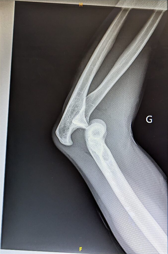
- AP and lateral radiographs before and after reduction
- Drop sign, ulnohumeral distance over 4 mm, indicates residual instability
- CT with 3D reconstruction for coronoid and radial head fracture patterns
- Wrist and forearm radiographs to exclude Essex Lopresti injury
- MRI for ligament injury in chronic instability
- Examination under anaesthesia with fluoroscopy to assess stability
Differential Diagnosis
- Simple elbow dislocation
- Isolated radial head fracture
- Monteggia fracture dislocation
- Distal humerus fracture
- Olecranon fracture
- Essex Lopresti injury
Treatment
Posterolateral Rotatory Instability
- Pathology
- Follows injury to the ulnar part of the LCL (varus extension stress)
- Allows transient rotatory subluxation of the ulnohumeral joint & secondary subluxation or dislocation of the radiohumeral joint
- Annular ligament intact thus the proximal radius & ulna move as one
- Clinical features
- Recurrent painful clicking/ snapping/ clunking or locking of elbow
- Occurs in extension 1/2 of arc with elbow in supination
- Trauma or surgery history usually present
- Radial head excision or Tennis elbow surgery often mentioned
- Lateral Pivot Shift Test (O‘Driscoll)
- Flexion of the elbow from extended position & supination of the forearm with valgus & axial load
- In posterolateral instability the radial head subluxes or dislocates posteriorly with rotatory subluxation of the ulnohumeral joint & palpably reduces at or just beyond 40 of flexion
- Radiographic Features
- Normal or slight widening of radiohumeral joint on AP XR
- Radial head posterior to capitellum on lateral XR (esp with supination)
- Fluoroscopy of pivot shift test will often reveal subluxation
- Treatment
- Often reconstruction required if symptomatic
- Tends not to become stable except possibly in the very early stages
- Technique
- Kocher approach
- The common extensor origin elevated to reveal origin of LCL on the lateral epicondyle
- Distally anconeus reflected posteriorly & ECU anteriorly
- Supinator crest of Ulna identified
- Capsulotomy & inspection of joint
- Anterior & posterior capsule plicated
- If able to reattach the LCL then performed
- If not then reconstructed with palmaris longus graft
- Isometric point located on the epicondyle & then fixed
- All sutures tied with the elbow flexed 30 & pronated
- Post op
- Cast for 4 weeks in elbow flexion of 90 & full pronation
- Then hinged splint with 30 extension stop for 6 weeks
- Then free splint for further 4-6 weeks
- Normal activity at 6 months
Fracture of Olecranon
- Type III fracture has ligamentous disruption
- If minimally comminuted then plate fixation stabilises the ulnohumeral joint (mandatory if fracture line in anterior half of notch)
- If coronoid involved then must be reduced & rigidly fixed
Fracture of Coronoid
- Most important part of the ulnohumeral articulation
- Resists posterior displacement of the ulna
- Classification
- Reagan & Morrey
- Type 1
- Small chip off tip & indicator of elbow dislocation and/or collateral ligament injury
- Stable & early mobilisation
- Type 2
- Up to 50% of coronoid elbow may be unstable
- If EUA has subluxation in less than 40-45 of flexion then need to stabilise
- If large enough then screw fixation performed
- If too small for screw then heavy braided sutures used (passed over coronoid fragment & tied through ulna)
- If still unstable then elbow distraction device used (eg. Compass Hinge)
- Type 3
- Greater than 50%
- Grossly unstable ulnohumeral joint
- Fix with screw & distraction neutralisation device
- If severely comminuted then realign with heavy suture & distraction device applied
Fracture of Radial Head & Coronoid with Dislocation
- Terrible triad
- Radial head replaced or fixed & coronoid fixed
- Protected with distractor
- Allows motion but eliminates the forces
Fracture of Radial Head with Attenuation or Tear of MCL
- 1-2% of patients with radial head fracture
- Classification of radial head fractures
- Mason (1954) three types
- Type I undisplaced
- Type II displaced wedge fragments
- Type III comminuted
- Type IV radial head fracture associated with elbow dislocation
- Added by Johnson
- Mason (1954) three types
- Principles of treatment
- Aim to retain the radial head as the secondary stabiliser to valgus stress by osteosynthesis
- If able to reconstruct then
- Stable arc within 40 of extension unrestricted motion after 2 weeks
- Dislocates with 60 of extension immobilise for 2 weeks then motion in hinged elbow splint with 30 extension stop for 2 weeks
- MCL not repaired
- Prerequisites for excision of fragment of radial head
- Excised fragment < 1/3 of radial head
- Excised fragment does not articulate with lesser sigmoid notch of ulna (ie anterolateral 1/3 of radial head this is also the site where fixation can be placed without impingement against the ulna during forearm rotation)
- If not able to reconstruct the radial head then
- Use of prosthesis or allograft
- Repair of the MCL acutely may be avulsed directly from the medial epicondyle requiring reattachment but if midsubstance then attachment more difficult
- Followed by locked hinged brace for 4 weeks then unlocked & motion in stable arc
- Total of 6 weeks
Fracture of Radial Head with Dislocated Elbow
- Mason IV injury
- If coronoid intact then reduce & assess the stability of the ulnohumeral joint
- Additional treatment according to fracture type
- Principles of treatment
- Type 1 (undisplaced)
- If arc of motion stable to 45-50 then place in splint with 60 extension stop for 10 days
- Full extension allowed in hinged splint for protection
- Type 2 (displaced)
- Need ORIF
- Will result in instability if radial head resected
- Usually not necessary to repair the MCL repair only if unstable arc of motion
- Type 3 (comminuted)
- Most difficult to treat
- If not able to reconstruct the radial head then excise & repair the collateral ligaments
- If still unstable then use of implant
- If still unstable then External Fixator allowing flexion an option (Compass Hinge from S & N)
- Allows distraction to maintain alignment but ROM allowed
- Removed at 3-4 weeks & splint used then
- Most difficult to treat
- Type 1 (undisplaced)
Chronic Elbow Instability
- Most activities only need 30-130 (100 arc) flexion-extension
- And 100 pronation-supination (50 each)
- Prerequisites for elbow stability
- Normal articular congruence
- Anterior band of MCL
- Ulnar part of LCL
- Annular ligament
- Resistance to valgus stress by radial head minimal when MCL intact
- Attenuated or torn ulnar part of LCL can cause PLRI with or without radial head
Chronic Non-Reduced Dislocation
- Normally 3rd world
- Secondary to fracture causing instability
- Operation indicated if painful
- Use external fixation distraction device
Recurrent Dislocation
- Rare
- LCL (PLRI) >> MCL
- 25% have lateral epicondyle nonunion
- LCL laxity most common
- MCL is unlikely to be lax
Posterolateral Rotatory Instability
- Secondary to LCL laxity
- Acute LCL tear after dislocation
- Tennis Elbow Release
- Describe clunk on flexion/ extension
- Posterolateral pain
- O’Driscoll Lateral Pivot Shift Test
- Treatment PLRI
- Kocher approach
- Drill holes x 2 base Tubercle of Crista Supinatoris
- Drill holes x 2 at Lateral Epicondyle
- Palmaris graft in figure of “8”
- Nestor 90% success
- MCL Deficiency
- Throwing injury
- Examination
- Valgus stress
- 40% Ulna nerve symptoms
- 40% Calcification MCL
- Pain at anterior bundle MCL on palpation
- Investigations
- Stress view
- Abnormal cf. opposite side if > 3mm difference
- Management
- Reconstruct with palmaris longus with two-strand technique
Complications
- Stiffness, with loss of terminal extension the commonest problem
- Heterotopic ossification
- Recurrent or residual instability
- Post traumatic arthritis
- Ulnar neuropathy
- Malunion or nonunion of the coronoid or radial head
- Capitellar wear from an overstuffed radial head arthroplasty
Prognosis
- Simple dislocations do well with early motion
- Complex injuries have higher rates of stiffness, arthritis and reoperation
- Functional arc is 30 to 130 degrees flexion with 50 degrees pronation and supination
- Most repaired terrible triad injuries regain a functional arc
- Missed anteromedial coronoid facet fractures lead to varus instability and early arthritis
- Chronic unreduced dislocations have poorer results than acute treatment
References
Reviewed by Professor Phong Tran, Head of Orthopaedic Surgery, Western Health. Last updated 10 October 2026.