
Question
A 19 year old footballer is tackled and lands on his shoulder. He has medial clavicle pain and difficulty swallowing.
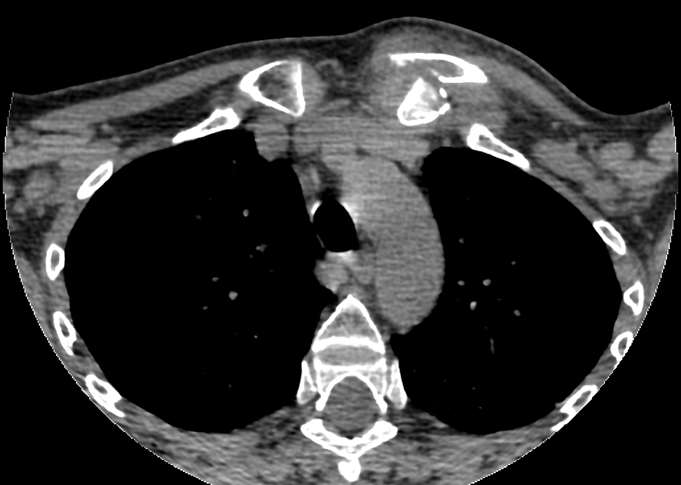
- What does the CT show and why does it matter?
- What other investigations are needed?
- Describe closed and open reduction.
Answer
CT findings
- Posterior displacement of the medial clavicle at the sternoclavicular joint
- Medial clavicle lies against the great vessels, trachea and oesophagus
- Dysphagia, dyspnoea, hoarseness and venous congestion signal compression
- In patients under 25 this may be a physeal fracture, the medial clavicular physis closes last (about 23 to 25 years)
Investigations
- Serendipity view radiograph (40 degree cephalic tilt)
- CT with contrast or CT angiography to assess vessels
- Chest examination for pneumothorax
Reduction
- Closed reduction under general anaesthesia within 48 hours
- Cardiothoracic surgeon available in theatre
- Supine with a bolster between the scapulae, traction in abduction and extension
- Towel clip on the medial clavicle to pull it forward
- Open reduction if closed fails or if chronic
- Stabilise with figure of eight semitendinosus graft or suture fixation
- Never use K-wires across the joint, migration into the mediastinum can be fatal
Related pages
Author Contributions
Orthofracs team
Reviewed by Professor Phong Tran, Head of Orthopaedic Surgery, Western Health. Last updated 10 October 2026.