
Question
A 35 year old man falls onto his outstretched hand. He has lateral elbow pain and a painful, limited range of forearm rotation.
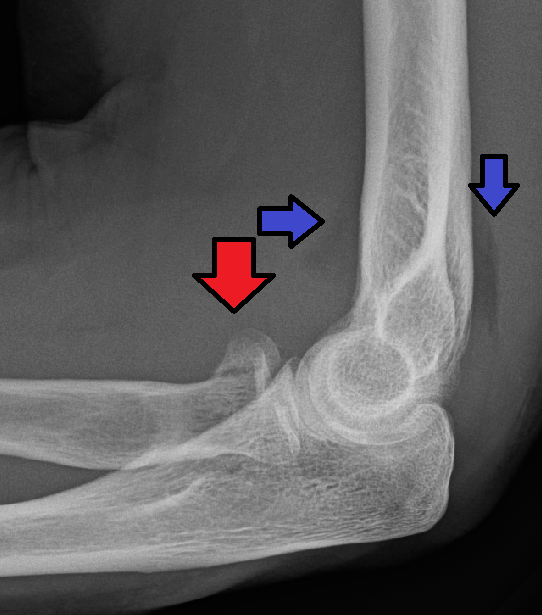
- Describe the radiograph.
- How do you classify the fracture and assess for a block?
- Outline management and associated injuries you would look for.
Answer
Radiograph
- Raised anterior and posterior fat pads indicate an effusion
- Radial head fracture line
- Radiocapitellar (Greenspan) view profiles the head
Classification
| Mason type | Description |
|---|---|
| I | Undisplaced |
| II | Displaced partial articular |
| III | Comminuted whole head |
| IV | With elbow dislocation (Johnston) |
- Aspirate the haemarthrosis and inject local anaesthetic
- Then test rotation for a mechanical block
Management
- Undisplaced or no block, sling and early motion
- Displaced partial fractures with a block, ORIF in the safe zone
- Comminuted fractures with more than three fragments, radial head replacement
- Excision only in low demand patients with stable elbow and forearm
Associated injuries
- MCL and LCL injury
- Coronoid fracture (terrible triad)
- Capitellar fracture
- Essex-Lopresti injury, palpate the DRUJ and interosseous membrane
- PIN injury with the Kocher approach, pronate the forearm
Related pages
Author Contributions
Orthofracs team
Reviewed by Professor Phong Tran, Head of Orthopaedic Surgery, Western Health. Last updated 10 October 2026.