
Question
A 72 year old man with type 2 diabetes has a stiff back. His lateral radiograph is shown.
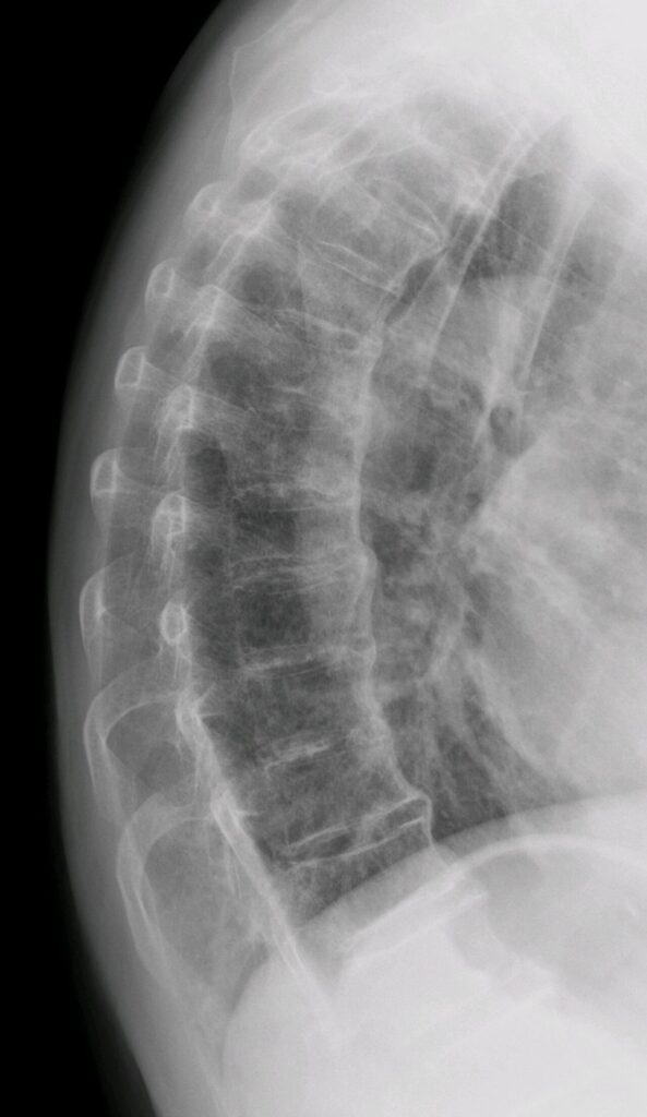
- What is the diagnosis and what are its criteria?
- How does it differ from ankylosing spondylitis?
- Why is it relevant after trauma?
Answer
Diagnosis
- Diffuse idiopathic skeletal hyperostosis (Forestier disease)
- Resnick criteria are flowing anterolateral ossification across at least four contiguous vertebrae, preserved disc height and normal facet and sacroiliac joints
- Associated with diabetes, obesity and metabolic syndrome
- Cervical osteophytes can cause dysphagia, and heterotopic ossification is common after hip arthroplasty
Comparison with ankylosing spondylitis
| Feature | DISH | Ankylosing spondylitis |
|---|---|---|
| Age | Over 50 | Young adults |
| Ossification | Thick, flowing, right sided anterior | Thin marginal syndesmophytes |
| Sacroiliac joints | Normal | Sacroiliitis |
| Facet joints | Spared | Fused |
| HLA B27 | No association | Strong association |
Trauma
- A long lever arm lets minor falls cause unstable extension fractures
- Fractures are easily missed on radiographs, so CT the whole spine
- MRI for epidural haematoma and cord injury
- Treat as unstable with long segment posterior fixation and keep the pre injury alignment
Related pages
Author Contributions
Orthofracs team
Reviewed by Professor Phong Tran, Head of Orthopaedic Surgery, Western Health. Last updated 10 October 2026.