
Orthopaedic Infections
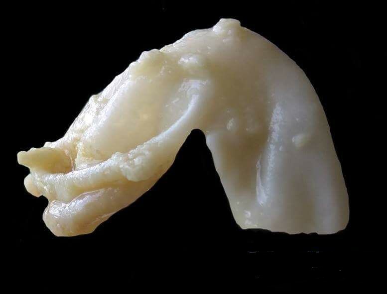
Hydatid Disease
- Echinococcus is the tapeworm
- Bone lesions are rare
- The larvae are ingested with the faeces of infected animals & are spread via portal circulation
- Therefore they settle in the liver & form Scolices
- These can rarely spread to bone with a predilection for
- Vertebrae
- Pelvis
- Femur
- Ribs
- Presentation may be incidental or with a pathological fracture
- Rarely they cause local pain & swelling
- XR shows the typical multiloculated bone cysts with mild expansion of cortices
- Serological testing for the diagnosis
- Casoni’s complement fixation test
Treatment
- Albendazole
- Surgery
- Curettage & bone grafting, with “sterilisation” of the cavity
Reviewed by Professor Phong Tran, Head of Orthopaedic Surgery, Western Health. Last updated 10 October 2026.