
Definition
- Most common 4 – 8 years of age
- Male: female 4-5: 1
- Bilateral in 10-12%
- No evidence that it is inherited
- Children are often small for their age
- Has been associated with ADD
Western Health Orthopaedic Registrar presentation – Legg-Calve Perthes Disease by Dr David Slattery
Presentation by Dr Praveen Kumar
Aetiology
- Idiopathic interruption of blood supply to the capital femoral epiphysis
- Associations with thrombophilia, passive smoking, low socioeconomic status and delayed bone age
Pathology
- Waldenström stages are initial, fragmentation, reossification and healed
- Lateral extrusion and collapse lead to aspherical head and hinge abduction
Classification
Herring lateral pillar (fragmentation stage)
| Group | Description |
|---|---|
| A | Lateral pillar height fully preserved |
| B | Over 50% of lateral pillar height preserved |
| B/C border | Narrow or poorly ossified pillar near 50% |
| C | Under 50% of lateral pillar height preserved |
Original publication Herring JA, Neustadt JB, Williams JJ, Early JS, Browne RH. The lateral pillar classification of Legg-Calvé-Perthes disease. J Pediatr Orthop. 1992;12(2):143-50.
- Gage sign, lateral calcification, lateral subluxation, horizontal physis and metaphyseal cysts
Stulberg (at maturity)
| Class | Description |
|---|---|
| I and II | Spherical head, low arthritis risk |
| III and IV | Ovoid or flat congruent head |
| V | Flat head with incongruent joint, highest arthritis risk |
Original publication Stulberg SD, Cooperman DR, Wallensten R. The natural history of Legg-Calvé-Perthes disease. J Bone Joint Surg Am. 1981;63(7):1095-108.
Clinical Features
- Limp with mild pain, often referred to the knee
- Loss of abduction and internal rotation
Investigations
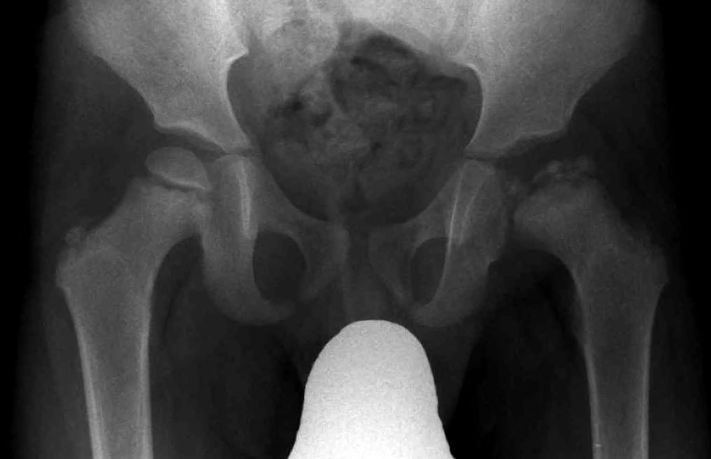
- AP and frog lateral pelvic radiographs
- MRI or bone scan in the early stage
Management
- Aim to contain the femoral head and preserve ROM
- Onset under 6 years, observation and physiotherapy
- Onset over 8 years with pillar B or B/C, femoral or pelvic osteotomy improves outcome (Herring 2004)
- Hinge abduction treated with valgus extension osteotomy or shelf acetabuloplasty
Prognosis
- Worse with older onset, female sex, pillar C and head at risk signs
- Stulberg class predicts adult osteoarthritis
Reviewed by Professor Phong Tran, Head of Orthopaedic Surgery, Western Health. Last updated 10 October 2026.