
Anatomy
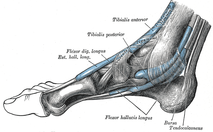
- Tibial nerve beneath the flexor retinaculum, posteroinferior to the medial malleolus
- Contents from front to back are tibialis posterior, FDL, posterior tibial vessels, tibial nerve and FHL
- Nerve divides into medial and lateral plantar and medial calcaneal branches
Aetiology
- Space occupying lesions, ganglion, varicosities, lipoma, accessory muscle
- Hindfoot valgus and trauma
- Tenosynovitis
- Idiopathic in a large proportion
- Systemic causes such as diabetes, hypothyroidism and inflammatory arthritis
- Anterior tarsal tunnel syndrome is compression of the deep peroneal nerve on the dorsum of the foot
Clinical Features
- Burning pain and paraesthesia in the sole, worse at night
- Positive Tinel sign over the tunnel
- Dorsiflexion and eversion test reproduces symptoms
- Symptoms worse with standing and walking
- Medial heel numbness when the calcaneal branch is involved
- Weakness of the intrinsic muscles is a late sign
Investigations
- MRI or ultrasound for a mass
- Nerve conduction studies are less reliable than in carpal tunnel syndrome
- Weight bearing radiographs to assess hindfoot alignment
- Exclude lumbar radiculopathy and peripheral neuropathy
Management
- Orthoses, NSAIDs and injection
- Release of the flexor retinaculum and the abductor hallucis fascia over the plantar branches
- Best results when a compressive mass is found
- Correct hindfoot valgus with orthoses
- Revision release has poorer outcomes
- Complications include wound breakdown and persistent symptoms
Differential Diagnosis
- Plantar fasciitis
- S1 radiculopathy
- Diabetic peripheral neuropathy
- Baxter nerve entrapment
Reviewed by Professor Phong Tran, Head of Orthopaedic Surgery, Western Health. Last updated 10 October 2026.