
Anatomy
Gastrocnemius
- Origin: via two heads.
- lateral head
- arises from distal femoral epiphysis, on lateral surface of lateral condyle, by tendon that leaves smooth pit above popliteus
- medial head
- arises via tendon from smooth shallow pit on medial condyle at distal end of supracondylar ridge, & via fleshy fibres from popliteal surface of femur
- medial & lateral heads form medial & lateral borders of distal half of popliteal fossa
- They converge to lie side by side, with deep surface covered by aponeurosis which rubs on soleus aponeurosis
- lateral head blends with soleal aponeurosis distally, but medial head is separate, & has tendon of plantaris interposed between it & soleus fascia
- lateral head
- Insertion:
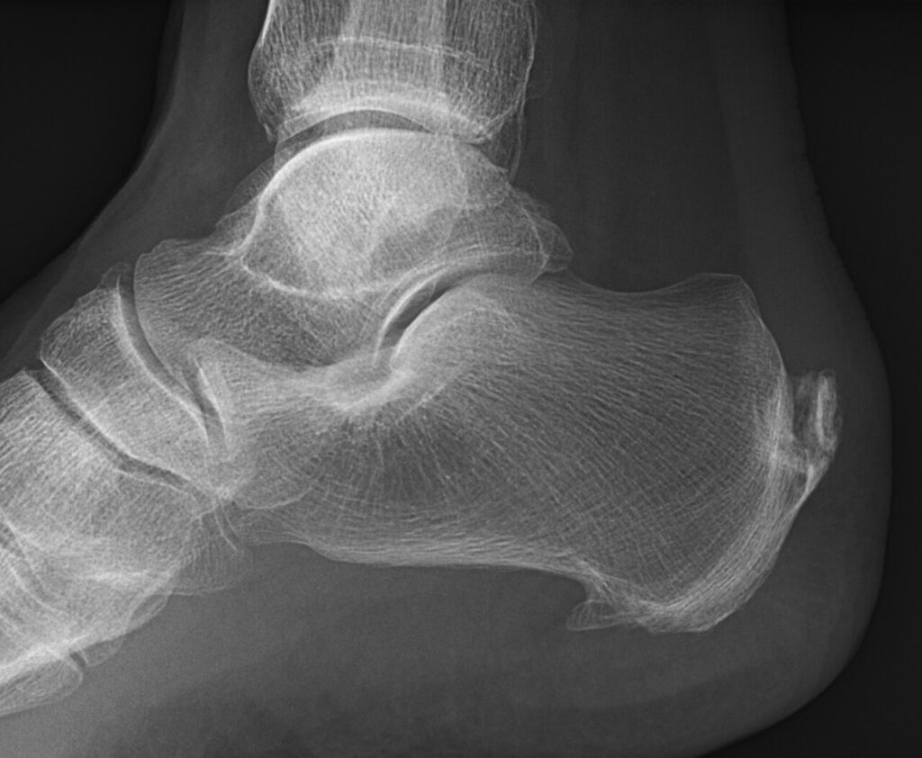
- tendon of insertion rotates through 90°, (DAMP), & inserts into middle third of posterior surface of calcaneus, separated from it by bursa. spiraling of tendon helps in storing energy
- Innervation
- tibial nerve (S1, S2).
Plantaris
- This vestigial muscle is seen in 93%
- Origin
- above gastrocnemius on lateral supracondylar ridge & its thin, twisted ribbon of tendon runs down between soleus & medial head of gastroc to insert into common tendon
- Innervation
- tibial nerve (S1, S2)
Soleus
- Origin:
- Arises from tibia (mostly) & fibula.
- fibular origin
- upper fourth, including head, then there is fibrous arch which spans tibial NV bundle (much like FDS) to reach tibia
- tibial origin
- middle third, down to hands breadth below lower end of soleal line
- fibular origin
- muscle is flat with deep & superficial aponeurosis; between aponeuroses short thick fibres run from anterosuperior down & posteriorly, & tendon of insertion arises from posterior layer
- On anterior surface of deep aponeurosis is slender bipennate muscle belly attached to centre of aponeurosis. This belly lies on NV bundle, in groove between FDL medially & FHL laterally
- muscle fibres of soleus contain deep venous plexus that can give rise to DVTs
- Arises from tibia (mostly) & fibula.
- Innervation:
- two separate branches of tibial nerve (S1, S2)
Structure of Achilles Tendon
- mostly type I collagen,
- < 4% strain
- no damage
- 4-8% strain
- microscopic damage
- > 8% strain
- macroscopic rupture occurs
- < 4% strain
- Type III collagen is found post rupture, & is not as strong
Blood Supply of Achilles Tendon
- controversial
- density of blood vessels is lower in middle 1/3 of tendon
- Blood flow decreases with increasing age
Reviewed by Professor Phong Tran, Head of Orthopaedic Surgery, Western Health. Last updated 10 October 2026.