
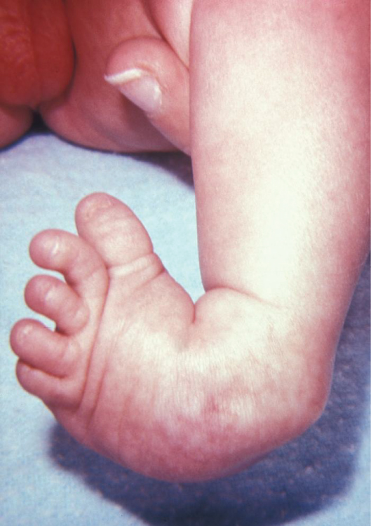
Definition
Congenital deformity of the foot with
- Equinus & varus deformity of heel
- Forefoot adduction
- Midfoot supination
Epidemiology
- Commonest of the congenital orthopaedic abnormalities
- 1.5:1000 live births
- Males>Females – 2:1
- 30-50% bilateral
- Much more common in Polynesian & Maori & lower in Asians
- Wynne-Davies suggests that has polygenic inheritance
- Tachdjian suggests that
- Patient with CTEV that has one child affected then 25% chance of another affected
- If both parents are normal & have affected child then chance of another is 5%
Classification
- Idiopathic or Secondary
- Idiopathic
- Multifactorial
- Polygenic
- Environmental
- Multifactorial inheritance patterns
- 0.5% if one 2nd degree relative (aunt, uncle) has CTEV
- 2% if one parent has CTEV
- 5% if one child has CTEV
- 25% if one parent & one child has CTEV
- Environmental
- Uterine constriction (Oligohydramnios, Constriction Band)
- Drugs/ Chemicals (Aminopterin, Tubocurarine)
- Secondary
- Genetic
- Part of syndromes with Mendelian Inheritance
- Craniofacial dysplasia
- Diastrophic dwarfism
- Larsens syndrome
- Pierre-Robin
- Neuromuscular
- Associated with
- Spina Bifida
- Cerebral Palsy
- Arthrogryposis
- Muscular Dystrophies
- Spinal Muscular Atrophy
Pathogenesis
- Unknown at this stage
- Various theories
- Irani & Sherman 1972 – suggest abnormal cartilage anlage in anterior aspect of talus secondary to germ cell defect
- Isaacs in 1977 – found histochemical muscle abnormalities under EM but not detectable on EMG
- Atlas 1980 – found abnormal vascular abnormalities with ischaemic area in sinus tarsi region & this suggests an abnormality with blood supply to tarsal neck region
- Dietz 1983 – Posterior tibial tendon sheaths have significantly less cellular & cytoplasmic volume than those anteriorly
- Zimmy et al 1988 – found regional differences in the cellular nature of the fascia in clubfoot – medial fascia contained cells resembling myofibroblasts & mast cells
- Victoria-Diaz 1984 – there may be defect in tibial growth phase & so production of clubfoot deformity
- In summary » Postulated to be due to
- Primary germ plasm defect
- Cartilage anlage arrest
- Abnormal myofibroblasts
- Abnormal neuromuscular junction
- Retracting fibrosis
- Anomalous tendon insertions
- Ischaemia
- Packaging defect (oligohydramnios)
Pathology
- All tissues around the foot are abnormal
- Bones
- All the bones of foot abnormal
- No internal tibial torsion
- Femur, Tibia & the fibula especially often shorter
- Talus
- Head & neck deviated medially & plantarward
- Body rotated externally in the ankle mortise
- Body extruded anteriorly
- Smaller than normal
- Calcaneum
- In equinus
- Rotated medially
- Means that long axis of talus & calcaneum parallel
- Smaller than normal
- Navicular
- Subluxed medially towards the medial malleolus
- Cuboid
- Subluxed medially
- Forefoot
- Adducted & Supinated
- Cavus deformity may occur
- Lateral Malleolus
- Located posterior near the calcaneus
- Muscles
- Atrophy & contracture
- Triceps Surae
- Tibialis Posterior
- FHL
- FDL
- Tendon sheaths thickened around
- Tibialis Posterior
- Peroneals
- Ligaments & Fascia
- Shortening of
- Calcaneofibular ligament
- Posterior Talofibular ligament
- Deltoid
- Long & Short plantar ligaments
- Spring
- Bifurcate
- Plantar fascia
- Tendo Achilles with more medial insertion on the calcaneum » ↑ varus
- Joint Capsules
- Contracture of capsules of
- Posterior ankle joint
- Posterior subtalar joint
- Talonavicular joint
- Calcaneocuboid joint
Clinical Features
- Clubfoot
- 1. Deformity
- Heel equinus
- Heel varus
- Midfoot supination
- Forefoot adduction
- Maybe cavus
- 2. Features
- Curved lateral border of foot
- Devil’s thumbprint over the lateral malleolus
- Medial & Lateral skin creases
- Navicular fixed to medial malleolus
- Os calcis fixed to the lateral malleolus
- Heel small & high
- 3. General
- Calf atrophy
- Calf shortening
- Other Conditions
- Should all be excluded
- Spinal Dysraphism
- Arthrogryposis
- Neuromuscular Disorders
- When examining patient for first time assessment made of the degree of correction able to be achieved
- Mild
- Able to correct past neutral
- Postural Form of deformity
- 10% require surgery
- Moderate
- Correction within 20° of neutral
- Structural Form
- 50% require surgery at some stage
- Severe
- Correction to < 20° of neutral
- Severe structural abnormalities – Teratogenic
- 90% require surgery
Radiology
- Can assess prior to treatment with A-P & Lateral of foot
- Calcaneal & Talar ossification centres are present at birth with the cuboid appearing by 6 months
- Navicular will not appear until 2-4 years
- Simons CORR 1978 – suggested standard radiographic assessment
- Anteroposterior View
- Kite’s angle – Anteroposterior Talocalcaneal angle
- Ankle dorsiflexed 15° & tube at 30° from vertical
- Talocalcaneal angle normally 20-40°
- Less than 20° suggests the talus & calcaneum are becoming more parallel
- Suggested that with nonoperative management the correction of equinus should be delayed until the Kite angle is normalised to avoid breaking midfoot
- First Metatarsal-Tarsal Angle
- Line through the long axis of first metatarsal & the talus respectively allows evaluation of the degree of forefoot adduction
- Normal angle is 0-20°
- Lateral View
- Lateral Talocalcaneal Angle
- Angle formed by line drawn through the long axis of talus & line along the plantar aspect of the calcaneum
- Normally is 35-55°
- < 35° indicates hindfoot equinus
- Talocalcaneal Index
- Addition of the Talocalcaneal angles in A-P & Lateral
- Normally > 40°
Treatment
- Initially non operative & starts on Day One of life » “as the feet exit the birth canal”
- Aims of treatment
- Correct deformity early
- Correct fully
- Maintain in corrected position until foot stops growing
- Remember two types of club foot – Attenborough 1966
- Easy
- Resistant
- The resistant form identified by
- Thin calf
- Small high heel
- Medial crease
- Devil’s thumb print laterally
- (ie seen with Arthrogryposis)
- 1. Non operative Treatment
- The First Cast
- Correct the heel varus & forefoot adduction first – avoid breaking the midfoot
- One hand on knee & one on toes & manipulate into neutral position
- Then apply firm SLPOP over the holders fingers & apply knob at the end for ease of removal
- Moulding until sets
- Repeat these second daily until the forefoot adduction is neutral – usually at 1 week
- First LLPOP
- Heel equinus corrected with this
- SLPOP applied with gentle correction of equinus – push calcaneus upwards & heel downwards & avoid pushing on midfoot
- Then extend as LLPOP with knee flexion & slight ER
- Subsequent casts applied at 3 days then weekly depending on progress
- This is repeated until the forefoot adduction & heel equinus corrected beyond neutral – ie Overcorrect
- Usually at 3 week time
- The last cast applied in full correction for further 3 weeks
- Then strapping & manipulation by the physiotherapists up to 12 weeks
- Splintage
- Bivalved AFO (front-back boots) & Dennis-Browne bar used full-time until walking age
- Removed to learn walking
- Applied when resting only up to 18 months
- Review
- See serially up to 8 years
- Outcome
- 50% of the feet are corrected by 6 weeks at end of casting
- 90% of mild
- 50% of moderate
- 10% of the severe
- If no correction then surgery required & no further non op treatment until surgery
- 2. Surgical Treatment
- Indications
- Failure of serial casting at 6 weeks
- Failure of subsequent splintage & strapping
- Timing
- Variable timing in literature
- Probably average is 3-9 months
- Advantage of
- Prior to fixed bony deformity & contractures
- Prior to walking
- There is literature to support later surgery at one year as anatomy more able to be recognised
- Indications
- Goals
- Release of all soft tissue restraints to allow proper positioning of the tarsal bones
- Approaches
- 1. Turco
- Posteromedial incision – curved
- From the base of first MT above the posterior tubercle of calcaneus to the TA
- The disadvantages include
- Crosses medial skin creases
- Difficult to explore the plantar fascia
- Difficult to explore the posterolateral corner
- May need a separate lateral incision particularly in older child
- 2. Cincinnati
- Posterior U shaped incision from the navicular medially curved posteriorly in the skin crease & ending at the calcaneocuboid joint
- Lateral exposure available with this approach
- Disadvantages
- Fear of loss of the posterior skin flap
- Exposure of the plantar fascia difficult
- Difficult to expose the proximal TA
- 3. Norris-Carrol
- Two incisions performed
- Curved incision from centre of os calcis to the talonavicular joint
- Second incision halfway between the TA & the lateral malleolus
- The disadvantage is two incisions
- 1. Turco
- Surgical Procedures
- Identify the N-V bundle & protect
- Posteromedial Release
- Z lengthening of the TA
- Posterior capsulotomy of
- Ankle joint
- Subtalar joint
- Release the Posterior tibiofibular ligament & the Calcaneofibular ligament
- FHL – intermuscular recession if the hallux flexes when foot dorsiflexed
- FDL – Z-lengthening if the lesser toes flex when foot dorsiflexed
- The above done at end of procedure if required
- Often the above is all that required
- Medial release follows if there is persistent varus
- Z-lengthening of the Tibialis Posterior & release of sheath
- Follow to the navicular insertion & this is guide to T-N joint
- The capsule of the T-N joint released
- The superficial fibres of the Deltoid ligament & the Spring ligament released
- At this stage usually able to reduce the navicular – if not then lateral release often required
- Plantar Release
- Usually if resistant cavus
- Able to reflect the Abductor Hallucis
- Release the
- Knot of Henry
- Long plantar ligament
- Plantar Fascia
- FDB from the calcaneum
- The calcaneocuboid capsule
- Medial ST joint released
- Lateral Release
- Usually severe forms where complete correction of the forefoot adduction not correctable with medial release
- Release the
- Bifurcate ligament
- Calcaneocuboid capsule
- Interosseous ligament
- Allows the calcaneocuboid joint to reduce independently to the talonavicular joint & correct the forefoot adduction & supination of midfoot
- Supplemental Fixation
- K-wire fixation across the
- Talonavicular joint
- Calcaneocuboid joint
- Subtalar joint
- Maybe required to hold position
- Postoperative Care
- LLPOP – in equinus if Cincinnati to protect the skin
- Neutral if other incisions
- ROS & take out wires at 2 weeks
- Then cast until 6 weeks
- Splintage as per non op treatment then
- Results
- Ponsetti 1963 – 71% good or excellent results
- Turco 1979 – 84% good or excellent results with posteromedial release
- Recurrence/ Failure
- 15% of operated clubfoot
- Not really recurrence rather
- Undercorrection
- Failure to maintain correction
- Prevented by
- Explanation preop about need for ongoing care
- Adequate initial correction
- Postoperative vigilance
- Suspect early with tight TA & stiff ST joint
- Usually see
- Forefoot adductus
- Hindfoot varus
- Curved lateral border of foot
- Cavus
- Younger than 2-3 years then can repeat the release
- Difficult +++
- Relatively high risks
- Benefits ↓ with advancing age
- May need to add lateral release or lateral column shortening
- If > 5 years then may need bony procedures to realign the forefoot or os calcis
- Forefoot Adductus
- Dynamic
- Correctable passively
- Best seen in swing phase
- Correct with SPLATT
- Transfer the lateral 1/2 of the Tibialis Anterior to lateral cuneiform or cuboid (via drill hole & pull-through with button in sole of foot)
- Fixed
- Bony procedure required
- May require MT osteotomies or Heyman-Herndon release
- Best procedure is Cuboid Decancellation
- Lateral incision & release of the C-C joint & reduction of it
- Then remove wedge of bone from the middle of the cuboid
- Other procedures to shorten the lateral column include
- Dillwyn-Evans procedure
- Shorten anterior process of calcaneus
- Medial soft tissue release
- Lichtblau osteotomy
- Hindfoot Varus
- Correction with calcaneal osteotomy
- Oblique sliding osteotomy best
- Slide the calcaneum laterally
- Can also perform Dwyer
- Lateral closing wedge calcaneal osteotomy
- Salvage Procedure
- Triple arthrodesis
- Best option for failed or resistant clubfoot
- Especially. if child > 12 years
- Choices are
- Standard triple arthrodesis
- Lambranudi arthrodesis if fixed equinus
- Plantargrade foot achieved with 95% good results but progressive Osteoarthritis (Ponsetti)
- Talectomy
- Reserved for the resistant arthrogrypotic club foot
- Poor results
- Complications
- Neurovascular injury
- Loss of foot (10% have atrophic dorsalis pedis artery bundle)
- Undercorrection
- Overcorrection (esp with Cincinatti)
- Forefoot adductus
- Hindfoot varus
Reviewed by Professor Phong Tran, Head of Orthopaedic Surgery, Western Health. Last updated 10 October 2026.