
Definition
Plantar fasciitis is a common cause of heel pain. It is a frustrating disorder due to its resistance to treatment.
Aetiology
- Repetitive microtrauma at the plantar fascia origin on the medial calcaneal tubercle
- Obesity and prolonged standing on hard surfaces
- Reduced ankle dorsiflexion from gastrocnemius or Achilles tightness
- Running, especially with sudden increases in mileage or poor footwear
- Pes planus and pes cavus both associated
- Seronegative spondyloarthropathy in bilateral or atypical presentations
Epidemiology
- Most common cause of plantar heel pain in adults
- Peak incidence 40 to 60 years
- Common in runners
- Bilateral in up to one third of patients
Anatomy
- Plantar fascia extends from calcaneal tubercles to MTPs + bases of the toes. It covers the intrinsic musculature + NV structures
- Passive MTP dorsiflexion places the inelastic plantar fascia under tension thereby elevating + stabilising the longitudinal arch = windlass effect
- Tensile forces are concentrated at the calcaneal origin
Pathology
- Degenerative fasciosis with collagen breakdown and fibroblast proliferation, with few inflammatory cells
- Thickening of the fascia origin, often over 4 mm on US
- Windlass mechanism tensions the fascia with toe dorsiflexion and loads the origin
- Plantar heel spur lies within the flexor digitorum brevis origin
- Heel spurs are common in asymptomatic people and do not explain the pain
Classification
- No accepted classification system
- Described by duration, with chronic cases persisting beyond 6 to 12 months of non-operative care
History
- Morning + start up pain.
- Initial pain relief with walking, but after prolonged walking pain recurs
- Pain localised to plantar fascia origin at medial calcaneal tubercle
- Bone scan = ↑uptake at plantar fascia origin
- Risk factors
- repetitive stress, obesity, middle age
- cavus foot 2˚ ↓hindfoot + ↓midfoot motion
- pes planus + TA tightness
- no evidence
- acute injury,
- heel spurs,
- bone spurs form superior to the plantar fascia and not within
- shoe type,
- walking surface,
- occupation
Examination
- Point tenderness at the medial calcaneal tubercle
- Pain reproduced by passive dorsiflexion of the toes (windlass test)
- Silfverskiöld test for isolated gastrocnemius tightness
- Negative calcaneal squeeze test to exclude stress fracture
- Tinel sign over the tarsal tunnel and first branch of the lateral plantar nerve
- Assess foot posture and heel fat pad thickness
Investigations
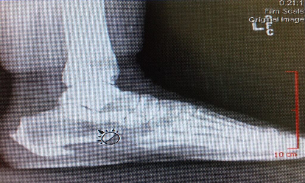
To exclude other pathology
- Numerous causes for subcalcaneal heel pain
- Bilateral
- ?seronegative arthropathy
- ankylosing spondylitis, Reiter’s
- ?seronegative arthropathy
- Neuropathic arthropathy
- diabetes + alcohol
- Vascular insufficiency
- Calcaneal stress #
- positive squeeze test = pain
- Bilateral
Differential Diagnosis
- Numerous causes for subcalcaneal heel pain
- Bilateral
- ?seronegative arthropathy
- ankylosing spondylitis, Reiter’s
- ?seronegative arthropathy
- Neuropathic arthropathy
- diabetes + alcohol
- Vascular insufficiency
- Calcaneal stress #
- positive squeeze test = pain
- Bilateral
Treatment
- No single method stands out as superior
- Stretching
- NSAIDs
- Heel cushion
- Steroid injection (no more than 2 per side)
- Casting. Success related to enforced rest?
- Surgery. No consensus on optimal procedure
Complications
- Heel fat pad atrophy after steroid injection
- Plantar fascia rupture after steroid injection or extensive release
- Lateral column pain and arch collapse after excessive release
- Injury to the first branch of the lateral plantar nerve or calcaneal branches
- Plantar heel wound problems and painful scars
- Persistent heel pain
Prognosis
- Self-limiting in most, with symptoms often lasting 6 to 18 months
- About 80 to 90% settle with non-operative care within 12 months
- Under 10% progress to surgery
- Releasing only the medial third to half of the fascia lowers the risk of arch collapse
Reviewed by Professor Phong Tran, Head of Orthopaedic Surgery, Western Health. Last updated 10 October 2026.