
Definition
- Congenital anomaly where-by an extra cervical rib or posteriorly fixed brachial plexus results in ↑ angulation of the subclavian artery & first thoracic nerve over the first rib
Aetiology
- Cervical rib from elongation of the C7 costal element
- Incomplete ribs often continue as a fibrous band to the first rib
- Other bony causes include anomalous first rib, long C7 transverse process and clavicle malunion
- Soft tissue causes include scalene hypertrophy and congenital fibromuscular bands
- Neck trauma and repetitive overhead activity
- Drooping shoulder posture
Epidemiology
- Cervical ribs occur in 0.4% of the population (70% are bilateral)
- Only symptomatic in 10% of cases
Anatomy
- Three sites of compression, interscalene triangle, costoclavicular space and subcoracoid space
- Interscalene triangle bounded by anterior scalene, middle scalene and first rib
- Brachial plexus and subclavian artery pass through the interscalene triangle
- Subclavian vein passes anterior to anterior scalene
- Lower trunk (C8 T1) lies on the first rib and is most often compressed
Pathology
- Neurogenic thoracic outlet syndrome accounts for over 90% of cases
- Lower trunk compression causes thenar wasting and ulnar sided sensory loss
- Arterial compression causes subclavian stenosis, post stenotic aneurysm, thrombosis and distal emboli
- Arterial cases nearly always have a bony anomaly
- Venous compression causes effort thrombosis (Paget Schroetter syndrome)
Classification
| Type | Description |
|---|---|
| True neurogenic | Objective neurological deficit |
| Disputed neurogenic | Symptoms and no objective findings |
| Arterial | |
| Venous |
History
- Pain and paraesthesia in the ulnar hand and medial forearm
- Symptoms worse with overhead activity or carrying loads
- Hand weakness and clumsiness
- Coldness, pallor and arm claudication suggest arterial involvement
- Swelling, cyanosis and heaviness suggest venous involvement
- Occupation and previous neck or shoulder trauma
Examination
- Supraclavicular fullness, tenderness or palpable cervical rib
- Wasting of thenar, hypothenar and interossei muscles
- Sensory loss in C8 and T1 dermatomes
- Adson, Wright hyperabduction and Roos elevated arm stress tests
- Compare blood pressure in both arms and listen for a subclavian bruit
- Exclude cervical radiculopathy and ulnar nerve entrapment
Investigations
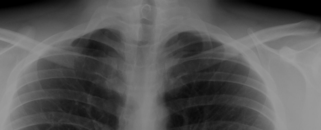
X-rays
- An occasional well formed rib is seen
- yet these are less likely to cause significant symptoms
- Elongation of the lateral mass of C7
- likely presence of a fibrous band
- this sharp fibrous band is more likely to result in symptoms
EMG
- not as good as clinical examination in establishing the diagnosis
Differential Diagnosis
- Carpal tunnel syndrome
- but the sensory & motor changes are not confined to the distribution of the median nerve
- Ulnar tunnel syndrome
- again the changes are not confined to the distribution of the ulna nerve
- Pancoast syndrome
- (apical carcinoma of the bronchus) may infiltrate the structures at the root of the neck causing pain numbness & weakness of the hand
- Clinically, large lump in the neck & chest X-Ray typical features of malignancy
- Cervical spine lesions
- eg disc prolapse or spondylosis where neck movements are limited
- TB & mets- X-Rays usually differentiate the cause
- Spinal cord lesions
- such as syringomyelia
- may cause wasting of the hand but other neurological features suggest diagnosis (dissociated pain & temperature loss)
- Cuff lesions
- have painful shoulder & arm movements
Treatment
- Nonoperative
- exercises to improve support of shoulder girdle associated with weight reduction are usually adequate
- Operative
- excision of the rib or fibrous band
- indicated if the above fails in the presence of severe symptoms
- significant neurological or vascular disturbances then
- excision of the rib or fibrous band
Complications
- Brachial plexus injury
- Long thoracic and phrenic nerve injury
- Pneumothorax
- Subclavian artery or vein injury
- Thoracic duct injury with chyle leak on the left
- Recurrence from scarring or incomplete resection
Prognosis
- Many neurogenic cases improve with physiotherapy and activity modification
- Surgical decompression relieves pain and paraesthesia in most patients
- Established muscle wasting seldom recovers
- Vascular cases do well with early decompression
- Disputed neurogenic cases have less predictable surgical outcomes
Reviewed by Professor Phong Tran, Head of Orthopaedic Surgery, Western Health. Last updated 10 October 2026.