
Video
Western Health Orthopaedic Registrar presentation – Cervical Spondylosis, Radiculopathy & Myelopathy by Dr James Churchill
Victorian Bone School Presentation – Cervical Spondylosis & Ankylosing Spondylitis
Definition
- Degeneration of lower cervical levels with loss of disc height, lipping of vertebral bodies (spondylophytes) & degeneration of intervertebral joints
Aetiology
- Age related disc degeneration with loss of hydration and height
- Repetitive loading and prior trauma
- Smoking
- Occupations with heavy loading or sustained neck postures
- Congenital canal stenosis predisposes to symptoms
- Genetic predisposition
Epidemiology
- More than 80% of the British population over 55 years have cervical spondylosis
Anatomy
- Seven cervical vertebrae and eight cervical nerve roots
- Cervical roots exit above the same numbered pedicle, the C6 root exits at C5 to C6
- Uncovertebral joints lie anterior and facet joints posterior to the foramen
- Normal sagittal canal diameter about 17 mm, under 13 mm is stenotic
- Pavlov ratio under 0.8 suggests congenital stenosis
- C5 to C6 then C6 to C7 most often involved
Pathology
- Encroachment
- Osteophyte formation from the:
- facet joints
- margins of the joints of Luschke
- spondylophytes from the
- vertebral body margin
- Osteophyte formation from the:
- Rarely osteophytes develop on the neurocentral lip which may encroach on the vertebral artery
- resulting in vertebral artery syndrome of
- dizziness
- vertigo
- tinnitus
- blurring of vision
- resulting in vertebral artery syndrome of
Classification
By clinical syndrome
- Axial neck pain
- Radiculopathy
- Myelopathy
- Myeloradiculopathy
Myelopathy severity by mJOA score
| Severity | mJOA score |
|---|---|
| Mild | 15 to 17 |
| Moderate | 12 to 14 |
| Severe | Under 12 |
History
- Patient usually over 40 years
- History
- Pain
- Complains of neck pain of gradual onset often worse in the morning
- Pain may radiate widely to the occiput, shoulder & arm
- Decreased Movement
- First movement to be lost is extension & may have marked limitation of lateral flexion when upright which improves on lying down
- Neurological
- Paraesthesia, weakness & clumsiness are occasionally features
- Pain
Examination
- May have tenderness of cervical musculature
- movements may be limited by pain
- very rarely may have signs of a cervical myelopathy with brisk reflexes in the lower limbs & ↑ tone
Investigations
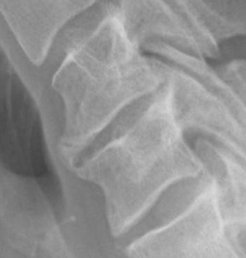
- X-ray
- Reduced disc height
- Cervical spondylosis (lipping of vertebrae & spondylophytes)
Differential Diagnosis
- Referred pain:
- Cervical disc degeneration (? not painful)
- Apical tumours (Pancoast syndrome, Horners & pain down the arm)
- Thalamic lesions (very uncommon)
- Thoracic outlet syndrome
- Local lesions:
- Carpal tunnel syndrome
- Neuralgic amyotrophy (brachial neuritis)
- Shoulder problems (AC joint, rotator cuff, Glenohumeral arthritis etc)
Treatment
- Nonoperative
- Rest analgesics & anti-inflammatories
- A cervical collar may be necessary at times of acute episodes
- Physiotherapy & local modalities
- Operative
- Decompression
- If associated with radicular symptoms or myelopathy may require decompression
- Fusion
- In some cases cervical fusion may be indicated in the absence of radicular symptoms
- Decompression
Complications
- Dysphagia and dysphonia after anterior surgery
- Recurrent laryngeal nerve palsy
- C5 palsy after decompression
- Oesophageal, vertebral artery or dural injury
- Retropharyngeal haematoma with airway compromise
- Pseudarthrosis and adjacent segment degeneration
- Post laminectomy kyphosis
Prognosis
- Restriction of movement usually persists but the discomfort resolves as time passes
- Many people have similar X-Ray changes with no or little discomfort at any stage in their life
- In dealing with predominant neck pain in the absence of neurological deficit & discrete radicular symptomatology the results of surgery do not significantly alter the natural history
Reviewed by Professor Phong Tran, Head of Orthopaedic Surgery, Western Health. Last updated 10 October 2026.