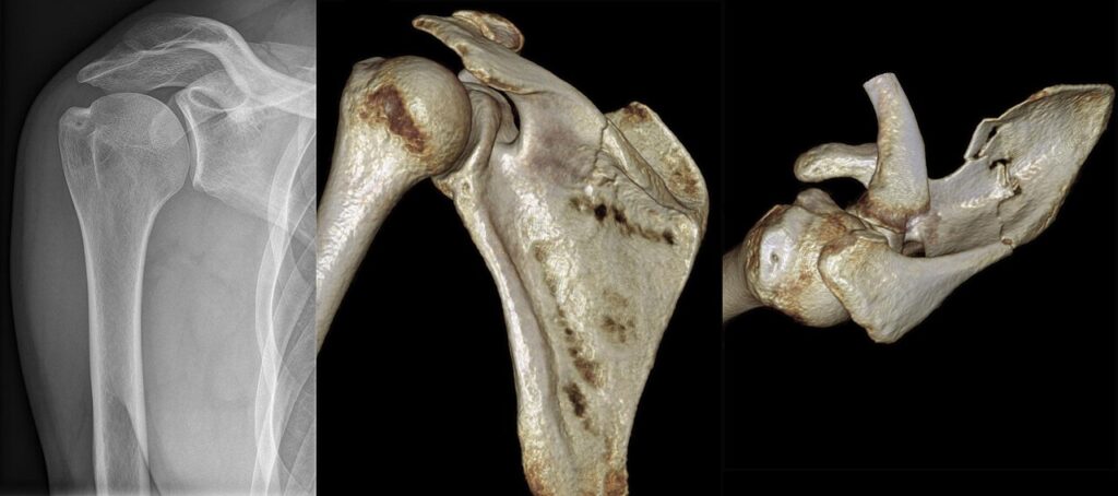
essential to rule out articular involvement with high quality AP view in which there is no overlap of humerus over glenoid
ideally, view should be purely tangential to glenoid
45 deg cephalic tilt allows evaluation of coracoid fractures
Apical Oblique View
CT Scanning
particularly helpful in evaluation of intra articular glenoid fractures
allows more accurate assessment of articular step off, as well as displacement & angulation of glenoid neck
Systematic Review
need to carefully assess entire scapular body & spine, acromion, coracoid, & glenoid
need to assess each articulation: glenohumeral, AC joint, & scapulo-thoracic
Treatment
Non-operative
vast majority of scapula fractures may be treated non operatively
closed reduction of these fractures is usually not possible
treatment consists of support of sling & early motion
most fractures will heal by 6 weeks
Operative
Indications
Body
rare
Neck
Controversial
> 10mm medial displacement
> 40 deg angulation
Medialization of glenoid up to 1cm is well tolerated by most patients
Glenoid Rim
> 10 mm displacement
> 25% of joint surface & displaced
(due to likelihood of instability)
Glenoid fossa
subluxation & instability of humeral head
step off of 3-4mm
> 20% of joint involved
but dependent on patient’s occupation, age, activity level, physiologic status, and hand dominance.
Acromion
depressed acromion fractures that encroach on subacromial space & interfere with rotator cuff function
Surgical Approach
anterior rim fractures are approached anteriorly & posterior rim fractures are approached posteriorly
transverse glenoid fractures may be directly reduced through anterior approach & have fixation via percutaneously inserted screws from above (thru deltoid)
Anterior Approach in Fractures of Glenoid
for fractures of anterior & inferior margins of glenoid, deltopectoral approach may be chosen
reduction of inferior glenoid fractures can be difficult due to proximity of axillary nerve
osteotomy of coracoid may be necessary for improved exposure
reattach coracoid with 4.5 mm cortex screw & absorbable washer to avoid splitting of coracoid tip
Implants:
3.5mm cortex screws or 4.0mm cancellous screws as lag screws
1/3 tubular plate may be applied below glenoid to lateral border of scapula as buttress
Extra-articular Scapular Fractures
Treatment Considerations
with glenoid neck fractures, (articular surface is intact) fracture extends from suprascapular notch area across neck to lateral border of scapula
glenoid neck fractures are often displaced but intact clavicle & AC joint will limit displacement & provide stability
implant of choice is often contoured 3.5 pelvic reconstruction plate which is applied to posterior border of glenoid & lateral
border of scapula
Implants
1/3 tubular plate, 3.5 mm DCP, or LC-DCP for fixation of Clavicle
3.5 mm DCP or LC-DCP
contoured 3.5 pelvic reconstruction plate
4.0 mm cancellous bone screws as lag screws
Prognosis
body
good prognosis for healing
neck
good prognosis for healing
if “operative scapular fractures” is treated non operatively
patient may develop abductor weakness & subacromial impingement
outcomes:
Scapular neck fracture influence of permanent malalignment of glenoid neck on clinical outcome
J. Romero. Archives of Orthopaedic & Trauma Surgery. 1434-3916 Volume 121 Issue 6 (2001) pp 313-316
authors analysed effect of associated shoulder girdle injury on glenoid displacement & influence of glenoid malalignment on clinical outcome
19 patients with scapular neck fractures were reviewed clinically & radiologically at mean of 8 years after injury
none of them has developed nonunion of scapular neck, & only one showed radiological signs of mild degenerative joint disease
glenopolar angle (GPA), which assesses rotational malalignment of glenoid about anteroposterior
axis perpendicular to scapular plane on plain X-rays was measured less than 20° in six patients
3 of them had sustained associated clavicular fracture or AC joint dislocation
other 3 patients had permanent severe malalignment of glenoid neck in absence of associated shoulder girdle injury
5 patients with GPA less than 20° complained of moderate or severe pain
13 patients with mild or no glenoid rotational displacement or medial displacement alone
11 patients had no or mild pain, & only 2 had moderate or severe pain
5 patients presented with reduced activities of daily living, 4 of them had severe glenoid rotational displacement
loss of motion was found in only 2 patients, & both had severely displaced glenoid neck
Reviewed by Professor Phong Tran, Head of Orthopaedic Surgery, Western Health. Last updated 10 October 2026.