
Incidence
- peak in 3rd decade
- Review 520 cases
| Type | Number |
|---|---|
| I | 185 |
| II | 119 |
| III | 204 |
| IV | 4 |
| V | 7 |
| VI | 1 |
Anatomy
- has fibrocartilage intraarticular disc
- Usually degenerative by 4th decade
- Clavicle may lie superior normally
Stability of ACJ
- Coracoacromial ligs
- Primary restraint to sup translation
- Primary suspensory ligament of upper limb
- Trapezoid Ligament
- Arises anterolateral on coracoid
- Inserts trapezoid ridge anterolateral to conoid
- Almost horizontal in sagittal plane
- PRIMARY restraint to AXIAL compression
- Conoid Ligament
- Arises posteromedial to trapezoid
- Inverted cone
- Inserts conoid tubercle
- Lies vertically
- PRIMARY restraint to SUPERIOR & ANTERIOR translation
- ACJ Capsule
- Strongest superiorly
- As reinforced by acromioclavicular ligament
- Has incomplete fibrocartilage intra-articular disc arising from it
- Usually degenerates by 4th decade
- Deltotrapezial Fascia
- Dynamic stabiliser
Motion at ACJ
- Only small 58 degrees
- 40 degrees at sternocla jt
- Motion is at scapulothoracic rather than ACJ
Classification
Allman grades I to III (1967), Rockwood modified (1989)
Classification of acromioclavicular joint dislocation
| Type | Description | Ligament injury |
|---|---|---|
| I | ACJ sprain | Incomplete AC ligament injury |
| II | ACJ disrupted, CCL intact or sprained | Complete AC, incomplete CC |
| III | Rupture of ACJ and CCL (25 to 100% subluxation) | Complete AC, complete CC |
| IV | Into trapezius | Complete AC, complete CC |
| V | High dislocation, more than 1 x clavicle width (100 to 300% subluxation). Disrupted trapezius and deltoid, end of clavicle subcutaneous | Complete AC, complete CC |
| VI | Subcoracoid dislocation | Complete AC, complete CC |
Original publication Allman Fl Jr. Fractures and ligamentous injuries of the clavicle and its articulation. J Bone Joint Surg Am. 1967;49(4):774-84.
Aetiology
- Direct trauma
- Usually direct fall onto adducted shoulder
- Clavicle remains in Normal position
- Arm falls down
- magnitude of force determines injury severity
- Usually direct fall onto adducted shoulder
- Indirect trauma
- fall on flexed elbow or outstretched arm
Pathology
- AC ligaments provide horizontal stability, CC ligaments vertical stability
- Trapezoid ligament lies lateral and conoid ligament medial
- Injury progresses from AC ligaments to CC ligaments then deltotrapezial fascia
- Scapula and arm sag inferiorly, giving apparent clavicle elevation
- Type 4 displaces posteriorly through trapezius
- Type 6 subcoracoid displacement is rare
History
- History of Traumatic Event
- Age of patient
- ? physeal injury
- Previous treatment
- nonoperative
- at least 3 months before considering surgery
- previous surgical procedures
- nonoperative
- Symptoms
- Pain
- with forward elevation
- Instability
- Posterior headache (nuchae)
- Pain
- Associated injuries
- distal clavicular fracture
- Functional disabilities
Examination
- Look
- Feel
- tenderness
- Move
- ROM
- inability to lift arm
- pain with forward elevation and wing out
- inability to lift arm
- Strength
- ROM
- Special Tests
- Stability
- AP translation
- Superior – inferior translation
- Shoulder shrug
- reduction of the distal clavicle with shoulder shrug differentiates between Type III from type V (distal clavicle buttonhole through deltotrapezial fascia)
- Stability
Investigations
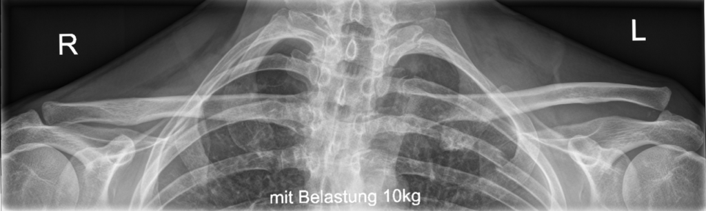
X-rays
Normal AC Joints
- 51% overriding clavicle
- 2% underiding
- 29% incongruent
- joint width 0.5-7 mm
Technique
- Zanca Views
- AP shoulder with 10° cephalad tilt
- 1/3 Penetration of shoulder X-ray
- Axillary, outlet and AP views of shoulder in scapular plane
- Cross arm adduction view
- AP shoulder
- measure clavicle override
- Stress views occasionally used (10lb weight)
MRI
- labral injury masquerading as ACJ pain
CT Scan
- nondisplaced lateral clavicle or acromion fracture
Differential Diagnosis
- Cervical spine disease: trapezial spasm
- Thoracic outlet syndrome
- Scapular dyskinesis
- Hyperlaxity
- Coracoid fracture
Treatment
Type I
- Nonoperative
- RICE
- Broad arm sling
- Avoid heavy stress & contact sport till Free Range of Movement & no pain to palpation
Type II
- Nonoperative
- RICE
- Broad Arm Sling 2/52
- Avoid heavy lifting, contact sports 810/52 to allow ligament healing
Type III
- Controversial
- Only 2 RCT OT vs NonOp
- Pt outcome 88% satisfaction Op or NonOp
- Further surgery 59% vs 6%
- Deformity 3% vs 37%
- ROM 86% vs 95%
- Only 2 RCT OT vs NonOp
- no indication for Operative reconstruction in literature to date
- Perhaps consider repair in
- Heavy labourer
- <25 yr undecided on career
Type IV, V, VI
- Operative
- Open Reduction and Internal Fixation
Chronic Symptomatic Grade III
- Poor results excision distal clavicle
- Convert long high riding clavicle to short high riding clavicle
- Must reconstruct lig as well
Specific Procedures
- Weaver Dunn Reconstruction
Complications
- Joint stiffness
- Deformity
- CC ligament and soft tissue calcification
- AC OA
- Associated fractures
- Distal clavicle osteolysis
Prognosis
- Weaver Dunn Reconstruction
Complications
- Joint stiffness
- Deformity
- CC ligament and soft tissue calcification
- AC OA
- Associated fractures
- Distal clavicle osteolysis
Reviewed by Professor Phong Tran, Head of Orthopaedic Surgery, Western Health. Last updated 10 October 2026.