
Indications
- Posterior wall and posterior column acetabular fractures
- Transverse and T-type fractures with mainly posterior displacement
- Irreducible or incarcerated posterior hip dislocation
- Pipkin femoral head fractures, via surgical dislocation
- FAI, labral and chondral surgery, and modified Dunn realignment for SCFE
Position
- Kocher-Langenbeck prone or lateral, hip extended and knee flexed throughout to slacken the sciatic nerve
- Ganz dislocation lateral decubitus with a sterile bag anterior to the table
Landmarks and Incision
- Landmarks are the PSIS, greater trochanter and femoral shaft
- Kocher-Langenbeck runs from near the PSIS to the trochanter tip, then down the shaft
- Gibson and Ganz use a straight lateral incision centred on the trochanter, slightly anterior
Internervous Plane
- Kocher-Langenbeck has none, splitting gluteus maximus (inferior gluteal nerve) in line with its fibres
- Gibson uses the interval between anterior gluteus maximus and TFL and gluteus medius (superior gluteal nerve)
- Gibson reflects all of gluteus maximus posteriorly, avoiding denervation of its anterior part
Superficial Dissection
- Fascia lata incised in line with the femur
- Gluteus maximus split proximally only to the first nerve branches to its anterior part
Deep Dissection
Kocher-Langenbeck
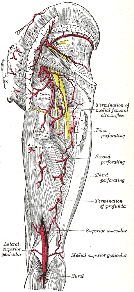
- Sciatic nerve identified on quadratus femoris
- Piriformis and conjoint tendon divided 1.5 cm from insertion to protect the deep MFCA branch
- Exposes the posterior column from greater sciatic notch to ischial tuberosity
Ganz surgical dislocation
- Trochanteric flip osteotomy keeps gluteus medius and vastus lateralis on the fragment
- Cut exits anterior to piriformis, which stays on the stable trochanter
- Z-shaped anterolateral capsulotomy stays anterior to the retinacular vessels and lesser trochanter
- Anterior dislocation in flexion and external rotation after dividing the ligamentum teres
Structures at Risk
- Sciatic nerve, especially with hip flexion or forceful retraction
- Deep MFCA branch, posterior to obturator externus and anterior to the conjoint tendon
- Superior gluteal neurovascular bundle at the greater sciatic notch
Extension
- Proximal extension limited by the superior gluteal bundle
- Trochanteric osteotomy improves exposure of the superior acetabulum and dome
Reviewed by Professor Phong Tran, Head of Orthopaedic Surgery, Western Health. Last updated 10 October 2026.