
Indications
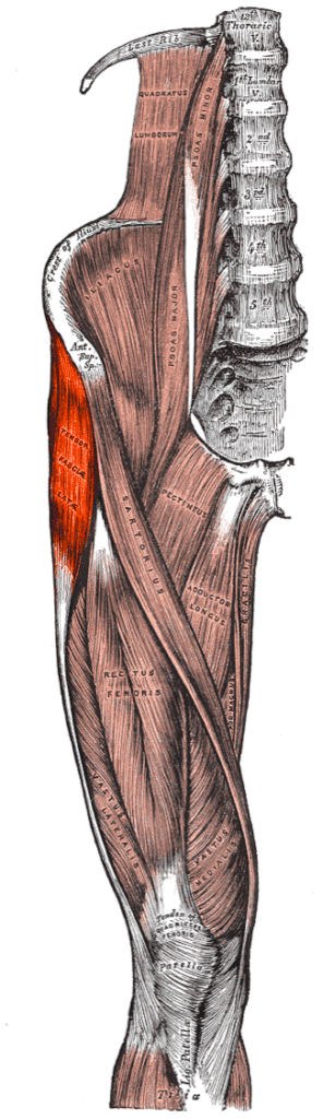
- Uses the interval between TFL and gluteus medius
- Basis of several minimally invasive anterolateral arthroplasty approaches
- Total hip replacement and hemiarthroplasty
- ORIF of femoral neck fractures
- Synovial biopsy and access to the anterior capsule
Position
- Supine with a bolster under the buttock, or lateral decubitus
- Ipsilateral buttock at the table edge falls away from the field
Landmarks and Incision
- Palpate the ASIS and greater trochanter
- Starts distal and lateral to the ASIS, passes over the anterior trochanter and runs down the shaft
Internervous Plane
- No true internervous plane
- TFL and gluteus medius are both supplied by the superior gluteal nerve
- The nerve enters TFL deep and posteriorly, so the plane is safe distal to its entry
Superficial Dissection
- Fascia lata incised posterior to TFL
- Interval between TFL anteriorly and gluteus medius posteriorly developed bluntly
- Gluteus medius retracted posteriorly to expose the capsule and gluteus minimus
Deep Dissection
- Fat over the anterior capsule cleared, reflected head of rectus femoris may be released
- Partial release of anterior medius and minimus fibres, or trochanteric osteotomy, improves exposure
- Capsule incised in a T or H shape, or excised
- Anterior dislocation with external rotation, extension and adduction
Structures at Risk
- Inferior branch of the superior gluteal nerve to TFL, with proximal dissection of the interval
- Femoral nerve, artery and vein from anterior acetabular retractors and medial dissection
- Ascending branch of the lateral femoral circumflex artery, crossing the interval and needing ligation
- Abductors damaged by retraction or partial release, causing limp
Extension
- Distal extension by splitting or elevating vastus lateralis to expose the shaft
- Proximal extension limited by the superior gluteal nerve
- Röttinger (OCM) variant uses the same interval through a shorter lateral incision
- OCM preserves the abductors entirely
Reviewed by Professor Phong Tran, Head of Orthopaedic Surgery, Western Health. Last updated 10 October 2026.