
Position
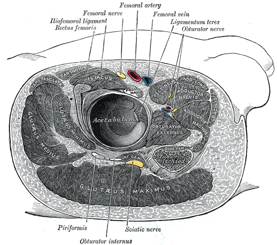
Supine (as First Described by Hardinge, K. (1982))
- buttock over hangs over edge of table
- tilt table away so patient lies flat
- prep & drape leg free
Lateral Decubitus
- Patient centred on table
- Posterior (Sacral) and anterior (ASIS) pelvic supports
- Hips and Knees flexed to 30 degrees (gel pads to protect bony protuberances)
- Prep & drape leg free
Landmarks
- ASIS
- Greater trochanter
- Shaft of femur
- Vastus lateralis ridge
Incision
- Flex the hip 30°
- Incision has the GT at its mid point longitudinally
- Extends 8cm parallel to the shaft of the femur along its anterior border
- Proximally it extends in a posterior direction ending at the level of the ASIS
Internervous Plane
- Intermuscular plane*
- Tensor fascia lata – Split lateral to innervation
- Gluteus medius – Split distal to innervation
- Superior gluteal nerve
- *No true Internervous plane as muscles share common innervation
Superficial Dissection
- Continue the incision through subcutaneous fat down to deep fascia
- The gluteal fascia and iliotibial band are exposed using blunt dissection and divided along the whole length and in the same direction as the skin incision.
- The tensor fascia lata is retracted anteriorly and the gluteus maximus posteriorly
Deep Dissection
- Detaching the abductor mechanism – if needed
- 1. Trochanter Osteotomy
- starting at Vastus Lateralis Ridge
- straight
- Chevron
- Use
- Gigli saw
- Osteotome
- Saw
- starting at Vastus Lateralis Ridge
- 2. Detaching Gluteus Medius
- Place a stay suture in the anterior portion of Gluteus Medius
- Identify the small prominence that lies at the uppermost end of the ridge of the vastus lateralis.
- Starting at this point and continuing to the APEX of the GT, the tendon of the Gluteus medius is incised using diathermy.
- Leave a cuff to be re-attached at closing.
- Extend the incision proximally between the fibres of gluteus medius.
- Do not extend more than 3-5 cm above greater trochanter to prevent injury to superior gluteal nerve
- Dissect off the Gluteus minimus tendon and the ligament of Bigelow
- Adduct and externally rotate the thigh
- 1. Trochanter Osteotomy
- Exposes Hip Joint Capsule
- Incise the capsule radially around its circumference
Dangers
- Nerve
- Vessels
- Femoral artery & vein
- Profunda femoris artery
- Fracture
- Femoral shaft
- Acetabular floor
References
- HARDINGE K: The direct lateral approach to the hip. J Bone Joint Surg Br. 1982;64:17–19.
Reviewed by Professor Phong Tran, Head of Orthopaedic Surgery, Western Health. Last updated 10 October 2026.