
Question
A 19 year old man treated for acute lymphoblastic leukaemia in childhood has bilateral knee pain.
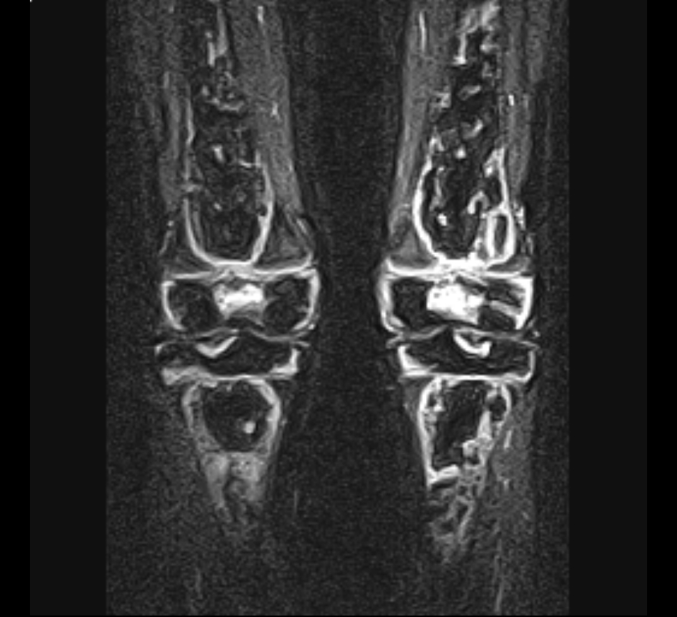
- Describe the MRI.
- Compare this condition with spontaneous osteonecrosis of the knee (SONK).
- Outline management.
Answer
MRI
- Multiple serpiginous geographic lesions in the distal femora and proximal tibiae
- Bilateral, metaphyseal and epiphyseal
- Secondary osteonecrosis after leukaemia treatment with high dose corticosteroids
Secondary osteonecrosis versus SONK
| Feature | Secondary osteonecrosis | SONK (subchondral insufficiency fracture) |
|---|---|---|
| Age | Under 45 | Over 60, women |
| Risk factors | Steroids, alcohol, sickle cell, Gaucher | Osteoporosis, meniscal root tear |
| Sites | Multiple, bilateral, femur and tibia | Single, weight bearing medial femoral condyle |
| Onset | Insidious | Sudden pain |
| Other joints | Hip, shoulder involvement common | Isolated |
Management
- MRI of both hips, as the hip is often involved
- Protected weight bearing and analgesia for small lesions
- Core decompression for pre collapse lesions
- Osteochondral allograft for focal collapse in young patients
- Arthroplasty for collapse with arthritis, using stems to bypass weak metaphyseal bone
Related pages
Author Contributions
Orthofracs team
Reviewed by Professor Phong Tran, Head of Orthopaedic Surgery, Western Health. Last updated 10 October 2026.