
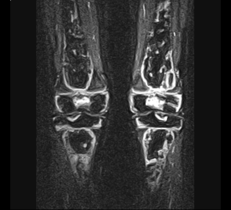
Osteonecrosis of the knee includes spontaneous osteonecrosis (SONK) in older adults and secondary osteonecrosis linked to steroids, alcohol and other risk factors. Secondary disease is often bilateral and multifocal, while SONK is usually a single medial femoral condyle lesion.
- Can be Idiopathic or Secondary
SONK
- Spontaneous Osteonecrosis Knee
- Aetiology
- Microtrauma
- Primary vascular ischaemia
Pathogenesis / Pathology
- Necrosis
- Inflammation/ Revascularisation
- Repair
- Remodelling
- ± secondary Osteoarthritis
- Initially localised depression of condylar surface
- Then articular sequestrum becomes partially separated as hinged flap
- May separate completely
- Result is crater with fibrocartilage base
Clinical Features
- Usually healthy woman age 60+ years
- Sudden onset of severe knee pain
- Almost always medial femoral condyle
- Exquisite local tenderness
- May be effusion
- Other side rarely involved
- SONK of tibial plateau less common
- Otherwise same as MFC
- May resolve spontaneously » allow course of non-op Treatment
Radiology
X-ray
- Initially normal
- Later develop
- Subchondral lucent line
- Crescent Sign
- Flattening of condyle
- Patchy sclerosis
- Can have rapid collapse into varus
Bone Scan
- Investigation of choice
- If have normal X-ray & painful knee in 60yr old think AVN & consider bone scan as may have degenerative meniscal tear at a/scope but don’t get better
- Focal ↑ in uptake
- One side of joint
- If both sides involved then more likely Osteoarthritis
MRI
- Discrete well defined low signal areas in subchondral region on T1
Classification
Modified Ficat and Arlet staging for the knee
| Stage | Radiographic features |
|---|---|
| I | Normal plain radiograph |
| II | Cystic or sclerotic lesion with normal bony contours |
| III | Crescent sign or subchondral collapse |
| IV | Joint space narrowing with secondary degenerative change |
Five stage radiographic staging (Insall)
| Stage | Radiographic features |
|---|---|
| 1 | Normal radiograph, positive bone scan |
| 2 | Subtle flattening of the weight bearing condyle |
| 3 | Radiolucent lesion with a sclerotic halo proximally and laterally |
| 4 | Subchondral collapse |
| 5 | Degenerative change with varus or valgus angulation |
- Extent is measured on the AP film
- A lesion wider than 50% of the condyle carries a poor prognosis and tends to deteriorate rapidly
Management
- Options are
- Initially watch to see if resolves
- A/S debridement & drilling
- Retrograde or Antegrade
- Bone graft or Osteochondral graft
- Valgizing HTO
- TKR if > 65yo
- See treatment algorithm below
Secondary AVN
- Much less common
- Pathogenesis
- Same sequence of events as SONK
- Caused by
- Steroid Therapy
- Alcohol
- Diver’s
- Marrow Proliferative Disorder
- Sickle Cell Disease
- SLE
Clinical Features
- Gradual onset of pain
- Lateral condyle in 60%
- Bilateral in 50%
- Is the natural history less predictable than hip AVN?
Radiology
- As for SONK
Management
- No standard management as uncommon
- Options as for SONK
| Feature | SONK | Atraumatic AVN |
|---|---|---|
| Age | Over 55 years | Often mid 30s |
| Site | Medial femoral condyle | Multiple areas |
| Laterality | 99% unilateral | 80% bilateral |
| Other joints | Knee only | Other joints involved in 60 to 90% |
| Location in bone | Juxta-articular | Epiphyseal, metaphyseal or diaphyseal |
- Treatment Results
- AVN may spontaneously resolve » allow nonoperative treatment first
- Arthroscope doesn’t appear to alter natural Hx
- Core Decompression
- Some success in stages 1 & 2
- Site of lesion not predictive of outcome
- Size of juxta-articular lesion
- Large lesion fared worse than medium or small both for tibia & femur
- But figures not recorded
- Role not as defined as AVN of hip
- Allograft experimental
- HTO
- Usually in younger high demand patient
- Koshino
- 37pts with 5yr follow-up
- Only 1pt with TKR
- Best when initially varus & lesion pre-drilled or grafted
- Arthroplasty
- Unicompartmental reasonable in SONK as virtually always medial comp only
- Marmor CORR 1993
- 89% good or excellent in 34 knees
- TKR
- Results not as good as for Osteoarthritis
- 85% success at 5yrs
- Mont, Hungerford et al Sept 2000 TKR successful in 60%
- 71% = 34 of 48 TKR successful at 9 years
- CONCLUSION
- INITIALLY WATCH AS MAY RESOLVE
- CORE DECOMPRESSION WORTHWHILE WITH GOOD OUTCOME
- TKR POOR RESULTS NOT UNLIKE AVN OF HIP
- ALGORITHM
- Stages I-III
- Non-Op 1st
- Persistent symptoms + no progression
- Core decompression
- Persistent symptoms + X-ray progression » TKR
- Initial presentation at Stage IV »TKR
Reviewed by Professor Phong Tran, Head of Orthopaedic Surgery, Western Health. Last updated 10 October 2026.