
Question
A 49 year old man has medial knee pain and stiffness. He injured the knee skiing a year earlier.
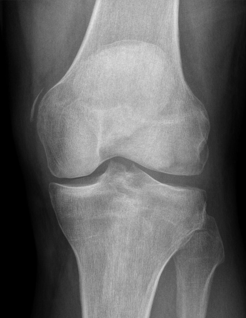
- Describe the radiograph.
- Describe the anatomy and grading of MCL injuries.
- How do you manage an acute MCL injury, alone and with an ACL tear?
Answer
Radiograph
- Ossification adjacent to the medial femoral condyle
- Pellegrini-Stieda lesion, calcification at the femoral MCL origin after injury
Anatomy
- Superficial MCL from medial epicondyle to the tibia about 6 cm below the joint
- Deep MCL (meniscofemoral and meniscotibial ligaments)
- Posterior oblique ligament
- Primary restraint to valgus at 30 degrees of flexion
Grading
| Grade | Opening at 30 degrees |
|---|---|
| I | Under 5 mm, firm end point |
| II | 5 to 10 mm, end point present |
| III | Over 10 mm, no end point |
- Valgus laxity in full extension implies cruciate and posteromedial injury
Management
- Isolated injuries heal well with a hinged brace and early motion
- Tibial side and Stener like lesions (sMCL above pes anserinus) heal poorly, repair them
- With ACL tear, brace the MCL for 4 to 6 weeks, then reconstruct the ACL
- Repair or reconstruct the MCL if valgus laxity persists in extension
- Pellegrini-Stieda lesions rarely need excision
Related pages
Author Contributions
Orthofracs team
Reviewed by Professor Phong Tran, Head of Orthopaedic Surgery, Western Health. Last updated 10 October 2026.