
Question
A 68 year old retired plumber has two years of night pain and stiffness in his dominant shoulder. External rotation is 5 degrees and his cuff strength is normal.
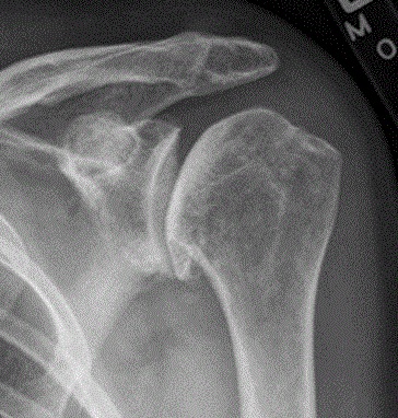
- Describe the radiograph.
- What further imaging would you request and why?
- What are the options, and how do you choose the implant?
Answer
Radiograph
- Joint space loss with an inferior humeral head osteophyte (goat’s beard)
- Subchondral sclerosis
- Humeral head centred under the acromion
- Primary glenohumeral osteoarthritis
Further imaging
- CT to assess glenoid version and posterior erosion
- Walch classification of glenoid morphology (A1, A2, B1, B2, B3, C, D)
- Assess fatty infiltration of the cuff (Goutallier)
- MRI or ultrasound if cuff integrity is uncertain
Options
- Analgesia, physiotherapy and steroid injection
- Arthroscopic debridement and capsular release in younger patients
- Anatomic total shoulder arthroplasty if the cuff is intact and the glenoid can be corrected
- Better pain relief and function than hemiarthroplasty
- Reverse shoulder arthroplasty for cuff deficiency, severe B2 or B3 glenoids, or age over about 70
- Augmented components or bone graft for posterior glenoid bone loss
- Loosening of the polyethylene glenoid is the main long term failure mode
Related pages
Author Contributions
Orthofracs team
Reviewed by Professor Phong Tran, Head of Orthopaedic Surgery, Western Health. Last updated 10 October 2026.