
Question
A 13 year old boy who plays soccer has activity related knee pain and intermittent swelling.
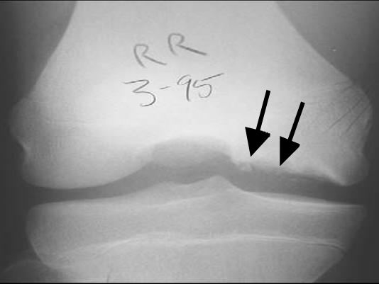
- What is the diagnosis and the usual site?
- What determines stability and prognosis?
- How is it managed?
Answer
Diagnosis
- Osteochondritis dissecans
- Lateral aspect of the medial femoral condyle is the usual site
- Wilson test reproduces pain with internal rotation as the knee extends
- Tunnel view shows the lesion best on radiographs
Stability and prognosis
- MRI signs of instability are a high T2 rim, cysts beneath the fragment and an articular breach
- Open physes give a much better chance of healing
- Smaller lesions in the classic site do better
Management
- Stable juvenile lesion, activity restriction for 3 to 6 months
- Failed or stable adult lesion, drilling
- Unstable fragment in situ, fixation with compression screws or bioabsorbable implants
- Unsalvageable fragment, removal and cartilage restoration such as microfracture, osteochondral grafting or ACI
Related pages
Author Contributions
Orthofracs team
Reviewed by Professor Phong Tran, Head of Orthopaedic Surgery, Western Health. Last updated 10 October 2026.