
Definition
- A condition whereby a segment of cartilage with subchondral bone separates from the articular surface
Incidence
- most common in the knee
- usually unilateral
- imost common cause of a loose body in the knee joint of a young person
- 15-20 knees / 100,000
- more common in males
- in the 2nd decade
Aetiology
- 2 schools of thought
- 1. due to an area of bone infaction with subsequent separation of a fragment
- 2. due to an osteochondral fracture which fails to heal
- this has been supported by
- occurrence of OCD especially in athletic young people
- associated with trauma to the joint e.g. direct blow, patellar dislocation
- production of OCD lesions in cadavic knees by applying axial compression & rotatory forces
- cadaveric studies showing that direct blow to the flexed knee can produce the classic OCD lesion at the lateral MFC
- this has been supported by
Associations
- significant valgus or varus deformity – 14%
- ACL deficiency – 7%
- patellar subluxation/ dislocation – 16%
History
- pain often activity related, usually of low intensity
Examination
- wasting,
- tenderness over the lesion,
- joint effusion
Wilsons Sign
- flex knee to 90°
- internally rotate tibia
- slowly extend the knee- in patients with OCD
- pain is produced at ~ 30° of flexion & is relieved with lateral rotation
Investigations
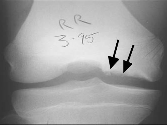
X-rays
- AP/ Lateral/Tunnel
- lesions most common on lateral aspect of MFC & posterior part of LFC
- rare on the patella & the trochlea
CT
- Defines lesion size, location and bony fragment integrity
- Useful when MRI is unavailable or contraindicated
- CT arthrography showing contrast beneath the fragment suggests instability
- Poor assessment of articular cartilage and the subchondral interface
- Radiation dose limits use in children
MRI
- Investigation of choice for lesion size, cartilage integrity and stability
- High T2 signal line beneath the fragment suggests instability
- Subchondral cysts, articular cartilage breach and fluid filled defect also suggest instability
- Instability signs are more reliable in adults than in juvenile lesions
- Detects loose bodies and craters from detached fragments
- Used to monitor healing during non-operative treatment
Arthroscopy
- Definitive assessment of cartilage integrity and fragment stability
- Probing detects softening, fissuring or a mobile fragment
- Guhl arthroscopic staging grades lesions from intact to detached with loose body
- Allows treatment at the same sitting with drilling, fixation or loose body removal
Natural History
- Children
- readily heals especially if on the posterior surface of the condyles
- Not all lesions spontaneously heal
- unstable
- lesions in the classical lateral MFC position
- less than 50% heal spontaneously
Treatment
- Depends on
- age of patient,
- stage of disease
- size & location of the lesion
Non-operative
- Indication
- if lesion is intact & nontender
- observation
- rest
- analgesia
- if tender
- splintage with R.O.M. exercises
- if lesion is intact & nontender
Operative
- indication
- if symptomatic despite nonoperative treatment
- displacement at any age
- persistence after physeal closure
- options
- if cartilage intact
- simple drilling of fragment with 1mm K- wire
- loose or sequestered lesions
- should be replaced & fixed
- if lesion small e.g. < 5mm can excise
- if cartilage intact
Prognosis
- incidence of Osteoarthritis higher
- in patients diagnosed & treated after skeletal maturity
- large fragment excised
Reviewed by Professor Phong Tran, Head of Orthopaedic Surgery, Western Health. Last updated 10 October 2026.