
Question
A 13 year old boy jams his finger in a car door. The finger is shown.
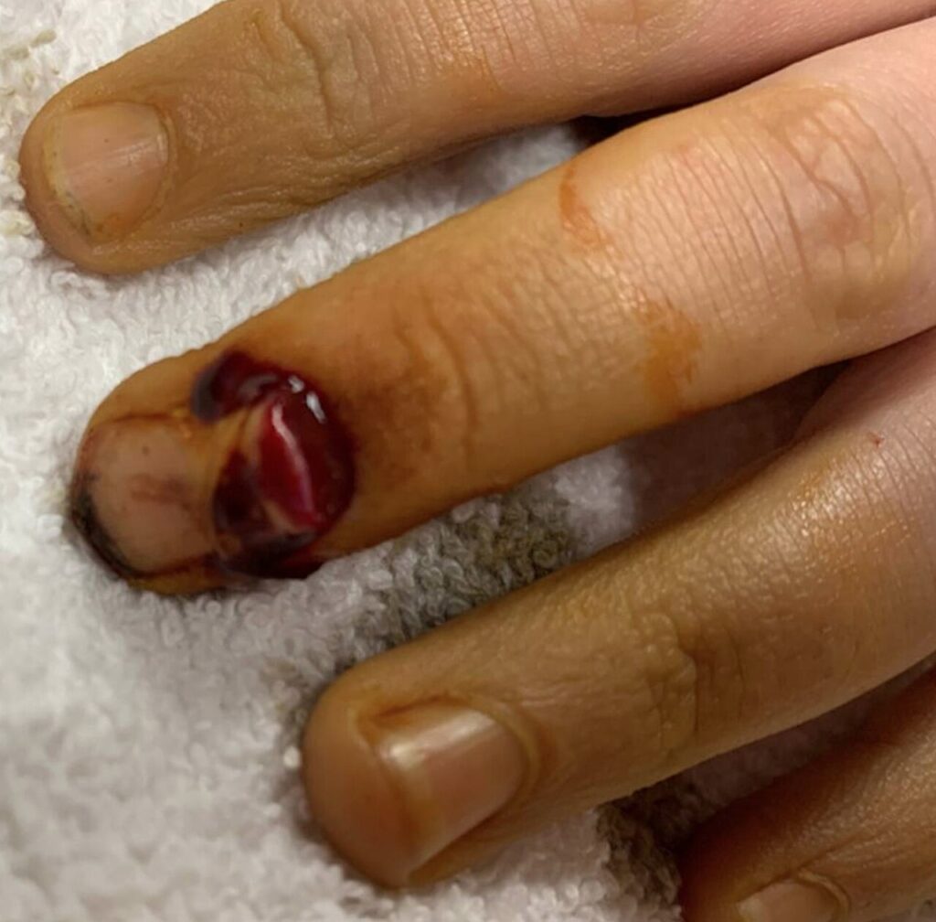
- What is the injury and why does it matter?
- What would a lateral radiograph show?
- Outline management.
Answer
Injury
- Seymour fracture, a juxtaepiphyseal or Salter-Harris I or II fracture of the distal phalanx with nail bed laceration
- Nail plate lies above the eponychial fold
- Open fracture, the nail bed is trapped in the fracture
- Looks like a mallet finger
Radiograph
- Flexion deformity at the distal phalangeal physis
- Widened physis on the lateral view
- Extensor tendon attaches to the epiphysis and FDP to the metaphysis, which flexes the distal fragment
Management
- Treat as an open fracture
- Remove the nail plate
- Extract the interposed nail bed from the fracture
- Irrigate and debride
- Reduce the fracture, K-wire if unstable
- Repair the nail bed with absorbable sutures and splint the eponychial fold
- Antibiotics (cephalosporin)
- Missed injuries cause osteomyelitis, nail deformity and growth arrest
Related pages
Author Contributions
Orthofracs team
Reviewed by Professor Phong Tran, Head of Orthopaedic Surgery, Western Health. Last updated 10 October 2026.