
Question
An 11 year old girl twists her ankle playing netball.
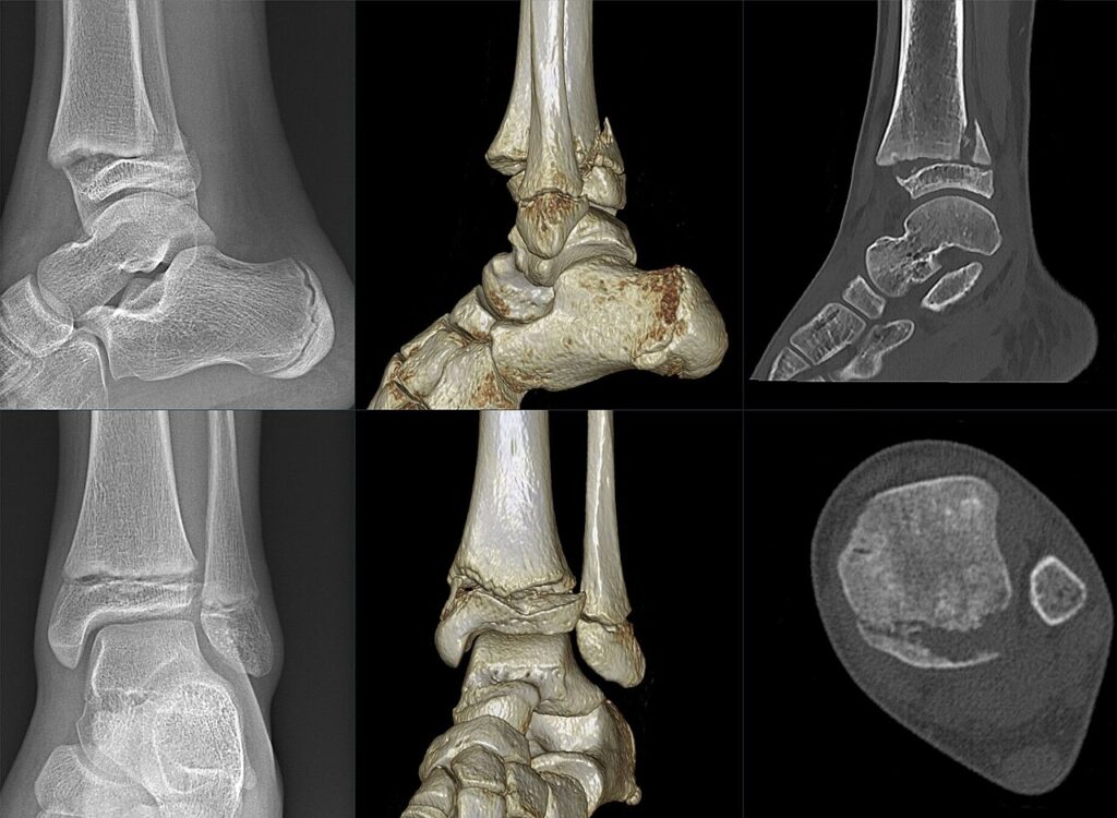
- Describe the imaging and classify the injury.
- What are the treatment goals?
- What complication should you warn about and how do you detect it?
Answer
Imaging
- Fracture through the distal tibial physis with a metaphyseal fragment
- Salter-Harris type II (Aitken I)
- Fibula intact or with a greenstick fracture
- CT defines displacement and excludes intra-articular extension
Treatment
- Undisplaced, above knee cast then below knee cast
- Displaced, closed reduction under anaesthesia
- Accept up to 2 to 3 mm residual physeal gap
- Gaps over 3 mm often contain interposed periosteum, open reduction and removal
- Fix with epiphyseal or metaphyseal screws parallel to the physis
- Avoid repeated manipulation
Complications
- Premature physeal closure in up to 40% of displaced SH II distal tibial fractures
- Residual physeal gap over 3 mm increases the risk
- Leg length difference and angular deformity
- Radiographs at 6 and 12 months
- Park-Harris growth arrest lines that are not parallel to the physis suggest a bar
- MRI or CT to map a physeal bar
Related pages
Author Contributions
Orthofracs team
Reviewed by Professor Phong Tran, Head of Orthopaedic Surgery, Western Health. Last updated 10 October 2026.