
Question
A 22 year old man has recurrent ankle sprains and lateral foot pain. His father has a similar foot.
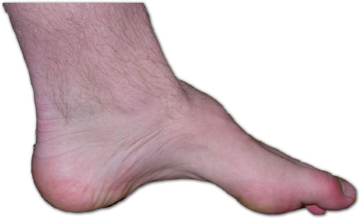
- Describe the foot and the likely underlying diagnosis.
- How do you examine the hindfoot?
- Outline surgical principles.
Answer
Foot and diagnosis
- High arch with a plantarflexed first ray and clawed toes
- Cavovarus foot
- Charcot-Marie-Tooth disease (HMSN type 1) in a young man with a family history
- Weak tibialis anterior and peroneus brevis, preserved peroneus longus and tibialis posterior
- Nerve conduction studies and genetic testing
Examination
- Peek-a-boo heel sign from the front
- Coleman block test, block under the lateral foot
- Heel corrects to valgus, hindfoot is flexible and forefoot driven
- Heel stays in varus, hindfoot is fixed
- Spine and neurological examination
Surgical principles
- Correct deformity, balance muscles and preserve joints
- Plantar fascia release
- Dorsiflexion osteotomy of the first metatarsal
- Peroneus longus to brevis transfer
- Lateralising calcaneal osteotomy if hindfoot is fixed
- Tibialis posterior transfer for foot drop
- Claw toe correction with Jones procedure for the hallux
- Triple arthrodesis for rigid adult deformity
Related pages
Author Contributions
Orthofracs team
Reviewed by Professor Phong Tran, Head of Orthopaedic Surgery, Western Health. Last updated 10 October 2026.