
Question
A 27 year old woman has deep ankle pain and swelling for six months after an inversion injury.
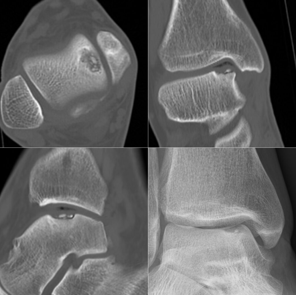
- Describe the imaging and the typical sites of these lesions.
- How are they classified?
- Outline management options.
Answer
Imaging
- Osteochondral lesion of the medial talar dome with a cystic subchondral component
- Medial lesions are posterior, deep and cup shaped, often atraumatic
- Lateral lesions are anterior, shallow and wafer shaped, usually traumatic
- MRI shows cartilage, oedema and fragment stability
Berndt and Harty
| Stage | Description |
|---|---|
| I | Subchondral compression |
| II | Partially detached fragment |
| III | Completely detached, not displaced |
| IV | Displaced fragment |
- Hepple MRI classification adds subchondral cysts (stage V)
Management
- Non-operative for stable lesions, protected weight bearing for 6 weeks
- Arthroscopic debridement and microfracture for lesions under 1.5 cm squared (about 107 mm squared) or under 15 mm diameter
- Fixation of large acute fragments
- Osteochondral autograft (OATS) for large or cystic lesions or failed microfracture
- Medial malleolar osteotomy may be needed for access
- Osteochondral allograft or matrix induced chondrocyte implantation for large defects
Related pages
Author Contributions
Orthofracs team
Reviewed by Professor Phong Tran, Head of Orthopaedic Surgery, Western Health. Last updated 10 October 2026.