
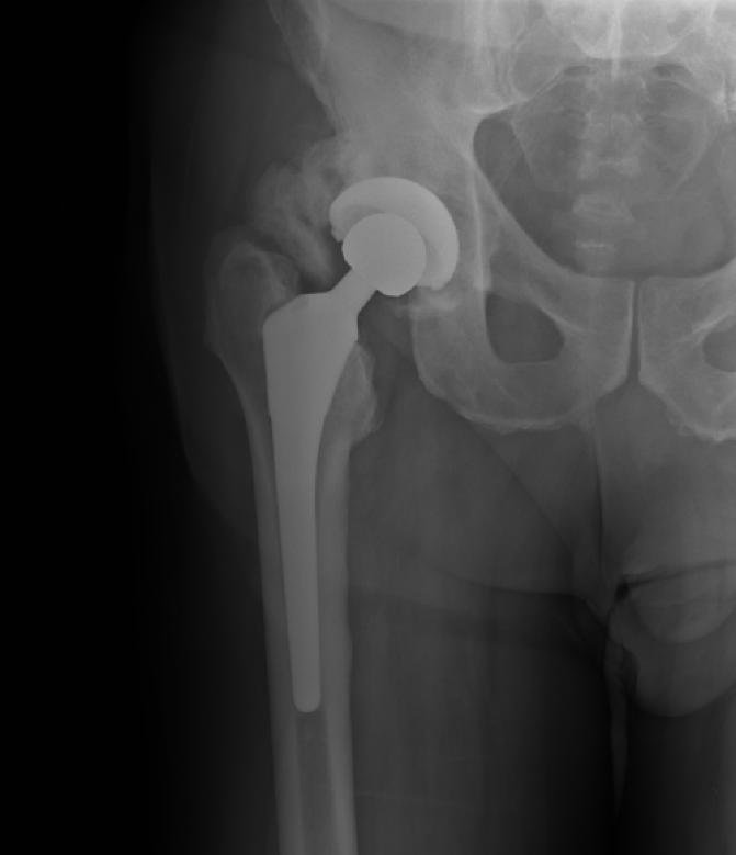
Definition
- Formation of mature bone outside the skeleton
- Different from ectopic calcification which consists of calcium salt deposits (mostly calcium phosphate) in tissues such as blood vessels, cornea & supraspinatus tendon. Relatively common in THJR (b/w 20 & 90 %)
Clinical Symptoms
Most asymptomaticPainful limitation ROM in up to 7 %
Aetiology
- Burns
- Head injury
- Direct trauma to soft tissues
- Total hip replacement
- Acetabular fractures & surgery
- Other articular injuries e.g. elbow
Pathology
- 4 characteristic zones in heterotopic bone
- innermost zone
- highly mitotic cells, possibly surrounded by dead muscle fibres
- hard to distinguish from sarcoma
- surrounding layers of cells
- producing osteoid
- zone of osteoblasts
- new bone with trabecular organisation
- may include osteoclasts modelling bone adjacent to host bone
- peripheral fibrous layer
- innermost zone
Pathogenesis
- process of bone formation is essentially the same as fracture healing
- Injury, haematoma, haematoma organised, converted to osteoid, bone
- ? reason -? role of BMP & other biologically active molecules
After Hip Replacement
- post THR incidence reported 8-90%
- only severe grades are symptomatic 1-33%
- Risk factors
- Previous HO on other side(up to 90 % risk in subsequent)
- Revision procedures
- Male sex
- ‘hypertrophic Osteoarthritis’
- trochanteric osteotomy
- DISH
Classification
Brooker classification, AP pelvis radiograph
| Class | Description |
|---|---|
| I | Isolated islands of bone in the soft tissues |
| II | Bone spurs with a gap of at least 1 cm between opposing bone surfaces |
| III | Near complete bridging with a gap under 1 cm |
| IV | Apparent ankylosis |
Original publication Brooker AF, Bowerman JW, Robinson RA, Riley LH Jr. Ectopic ossification following total hip replacement. Incidence and a method of classification. J Bone Joint Surg Am. 1973;55(8):1629-32.
Severe Type
- Fibrodysplasia ossificans progressiva (myositis ossificans progressiva)
- Rare condition
- Heterotopic bone mass replaces muscle tissue & restricts chest movement leading to early death
Differential Diagnosis
- bone tumour
Prophylaxis
- NSAIDs Schmidt et al
- No HO in 85 % of treated, 25 % of untreated
- None of HO in treated group was high-grade (III – IV)
- Dosage still under Ix
- 25 mg tds indocid for 6 weeks
- Maybe 2 weeks is adequate
- Beware usual side effects
- Radiotherapy
- Single dose 700 – 800 cGy within 1 – 4 days of surgery
- Or single preop dose of 800 cGy
- No secondary malignancy in study of 90 patients to 8 years
- Becoming more common prophylaxis because single shot & no compliance issues
Treatment
- Once formed, delay surgery at least 6 – 12 months before attempted excision
- Tc bone scan to assess activity
- Excision very effective in improving ROM
- Poor correlation between XR appearance & clinical ROM
- Pain relief following excision is variable
- NB: range of motion possible is, however, often significantly greater than one would expect from X-ray appearances alone. Many class 4 cases have an entirely functional range. The ankylosis is usually only apparent – remember the system is based on AP views only
- Excision can improve range of motion, but has less effect on symptoms
Reviewed by Professor Phong Tran, Head of Orthopaedic Surgery, Western Health. Last updated 10 October 2026.