
Definition
- Congenital disorder in which the 2 halves of the posterior vertebral arch (or several arches) fail to fuse
- Anencephaly = cranial end equivalent
- Multiple organ involvement due to abnormal innervation of structures supplied by affected part of nerve
Demographics
- Isolated laminar defects are seen in 5% of lumbar spine X-rays
- <1 per 1000 live births in Australia
- up to 5 per 1000 in some countries
- If one child is affected then there is a 10 fold risk for the next child
Aetiology
- Dysraphism
- Embryonic defect associated with maldevelopment of the neural tube & the overlying skin
- Environmental implications
- Folate deficiency
- Selenium deficiency in soil
- Maternal alcohol
- Maternal diabetes
- Fever at critical antenatal stage
- Usually occurs in the lumbar or lumbosacral region
- With neural involvement there can be motor, sensory & autonomic dysfunction
Pathology
Spina Bifida Cystica
- Severe dysraphism with absent laminae & prolapse of the theca ± its contents
- Visible Cyst Present
- Meningocoele
- CSF filled sac through the defect
- Cord & roots still confined within vertebral arches
- Myelomeningocoele
- Cyst including cord/ cauda/ nerve roots prolapsing
- Cyst lined by dura & arachnoid
- Commonest form
- Rachischisis
- Open Myelomeningocoele
- If the cord is still in its primitive state (ie. unfolded)
- neural plate forms the roof of the prolapsing sac
- Closed Myelomening
- If the neural tube is formed the roof of the sac is formed by dural membrane
Spina Bifida Occulta
- Midline laminal defect without prolapsing neural tissue
- Hidden defect
- There can be associated intraspinal defects
- Tethering of the conus
- Diastematomyelia
- Cysts or lipomata of the cauda
- There may be skin signs of this with a dimple, a pit, or a tuft of hair
Multisystem Involvement
Hydrocephalus
- ~ 72%
- Secondary to Arnold Chiari malformation ± tethering of the cord
- brainstem may be dragged distally, herniating through the foramen magnum
- obstructing outflow of CSF
- If untreated it will lead to cerebral atrophy & retardation
Neurological Deficit
- Myelomeningocoele is always associated with neurological changes at & below level of the lesion
- Even with occulta there can be neurology
- Many patients have upper limb neurology because of subtle neurological lesions
Visceral
- Bowel & bladder paralysis
- Tendency to trophic ulceration of sacral / buttock / feet skin
Clinical Features
- Assess early for level of lesion & annual full neuro check
- Clinical assessment of level of lesion is easier after 5 y.o.
Spina Bifida Occulta
- If there are skin signs then neurological deficit is more common
- May present at any age
- Usually partial cauda equina symptoms with bladder symptoms along with motor & sensory features
Spina Bifida Cystica
- Obvious saccular lesion at birth, that may have skin or just membrane covering
- An open myelomeningocoele will have neural elements merging with plum coloured skin around
- the periphery
- head may be enlarged with widened sutures due to hydrocephalus
- posture may suggest the neurological level
- associated abnormalities;
- hip dislocation
- genu recurvatum
- CTEV
- Claw toes
- Such deformities may be due to muscle imbalance or foetal positioning
- Neurological deficit;
- Is complete in a third
- Is complete at the level but there is some distal preservation in a third
- Is incomplete in a third
- 60% have upper limb neurological deficits (often require both hands free to perform simple tasks)
- Walking
- Thoracic – Most will not walk into adult life
- Lumbar – May continue walking if strong quads & no significant hip deformity
- Sacral – Useful walkers, often without orthoses in adult life
- Children with L-1 & L-2 neurosegmental levels are rarely community ambulators as adolescents
- but time spent walking in braces may be longer than in children with thoracic lesions
- so surgery to improve brace fitting should be considered
- Children with L-3, L-4, & L-5 neurosegmental levels with strong quads are usually good walkers.
- They usually require an ankle-foot orthosis to stabilise the ankle
- need for crutches or walking aids depends on the strength of abductors at hip
- Most children with sacral lesions are effective community ambulators until adulthood,
- when some deteriorate & cease walking. Hip deformity is uncommon
- but surgery is often necessary for foot & toe deformities
Investigations
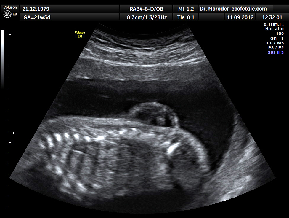
- Genetic counselling
- One affected » 1 in 25 risk for subsequent babies for neural tube deficits
- Two affected » 1 in 10 risk
- Three affected » 1 in 4 risk
- Screening using
- Maternal Alphafetoprotein levels in serum
- 16th to 20th week
- If high, then need USS ± amniocentesis for Diagnosis
- Ultrasound
- detects most anencephalics by 10 – 12 weeks on PV USS
- detects most by 16 – 18 weeks on standard abdo USS
- detects > 80% of spina bifida on standard USS at 18 weeks
- Amniocentesis
- Alphafetoprotein levels
- May still miss small skin-covered defects
- Maternal Alphafetoprotein levels in serum
- XR
- Midline bony defect
- Associated bony anomalies
- Unsegmented bars
- Hemivertebrae
- Fused rib s
- All of these can lead to abnormal curves
- MRI
- To visualise the cord & the lesion
- Brainstem herniation
- Diastematomyelia
- Syringomyelia
- Cysts/lipomata in the theca
Prevention
- Folate / multivitamins
- Some elect for antenatal Diagnosis » abortion
- If detected NTD » consider elective LUSCS at 36 weeks (less severe neurological defects than NVD at term)
Treatment
- Multidisciplinary in large centre
- Coordinator, neurosurgeon, orthopaedic, urologist, paediatric surgeon, psych, social worker, physio, orthotist, stomal, school aids etc
- For the focal deformity:
- Closure of skin over the defect within 48 hours
- For the hydrocephalus:
- Ventriculocaval shunt
- For the urogenital abnormalities:
- Permanent catheter or a diversionary procedure
- For the musculoskeletal problems:
- 2 important things to remember:
- the bones are weak – therefore forceful corrections may lead to Fracture
- the skin is insensate – therefore splinting must avoid pressure areas, & plasters are to be avoided
- Postoperative heterotopic calcification around hip may be problematic if high lesions
- 2 important things to remember:
Abnormalities & Their Management by Region
Spine
- Combination curves are common & can be severe
- Braces may slow the progression but many require surgical correction
- Kyphosis
- If present at birth » usually associated with hydrocephalus
- Usually lumbar
- Increase > 8o per year
- Correct if skin integrity compromised
- Scoliosis
- Most = paralytic (some are congenital or mixed)
- Most common in thoracic lesions
- Brace only for those with rapidly progressing curves & too young for surgery
- Indications for surgery
- Stable posture for standing & sitting
- Aim for level pelvis & trunk vertical on this
- Try to avoid need for hand support for sitting
- For stable fusion & prevent recurrence
- Skin integrity
- Respiratory function
- Most surgical corrections = >35o & 8 – 14 y.o
Hip
- 3 problems
- Deformity
- Dislocation
- Weakness (extension & abduction)
- Aim
- enable the child to stand straight in calipers & also to be able to sit in a chair
- Flexion
- Commonest deformity
- <20° & good walking potential » observe
- >20° & good walking potential » soft tissue release anterior & lateral
- Dislocation
- Reduction of dislocated hip does not improve walking potential
- May reduce to correct leg length discrepancy
- Usually associated with pelvic osteotomy to maintain reduction
- Weakness
- Above L1
- then all the muscles are flail
- splintage is all that is required
- Below L5
- deformity is one of flexion
- either a flexor muscle ‘slide’ on the ilium or a psoas tendon elongation can be performed
- Usually the lesion is between these levels
- imbalance leads to hip subluxation
- 50% of SB kids have subluxated or dislocated hips by the age of 2!
- In view of the dangers of splinting & plasters
- it is important to achieve muscle balance
- psoas tendon transfer from lesser to greater trochanter achieves this
- by decreasing flexor strength & increasing abduction & extensor strength
- (Sharrard advocates passing the tendon through a hole created in the ilium)
- (Mustard prefers to pass the muscle in front of the bone)
- As the child gets older it becomes more difficult to reduce the hip
- If the child is functionally OK then it is better to observe
- Above L1
Knee
- Aim
- knee that can be held straight in a caliper, & can also be flexed to sit in a chair
- Recurvatum can be treated with a careful quadriceps elongation
- Undeformed knee with Quads weakness = common
- Fixed Flexion
- Common with thoracic lesion
- < 20° can be observed
- >20° = if walker » posterior hamstring release ± capsular release
- Common with thoracic lesion
- Recurvatum
- Problematic > 6y.o
- Subcutaneous tenotomy of patella tendon
Tibia
- External torsion
- Often with ankle valgus
- Perform supramalleolar derotation osteotomy after 8y.o
- Internal torsion
- < 3 y.o. can perform osteoclasis of tibia & fibula
- Later stage = supramalleolar derotation osteotomy
Foot
- The aim is a plantargrade foot
- May be worth correcting deformity in non-walkers to look normal & wear normal shoes
- Varus
- Results in weight bearing on outer border of foot
- Surgical correction
- Valgus
- Need to determine if mortise or STJ origin
- Ankle
- Distal end of fibula more proximal than normal
- XR = growth plate more proximal than mortise
- Lower tibial physis is wedge-shaped
- Correct during growth by partial epiphysiodesis of tibial growth plate
- (Staples, Phemister, Closed drilling)
- Correct at later age by supramalleolar osteotomy
- STJ
- Usually controllable with footwear & orthoses until adolescence
- Os calcis osteotomy (medial based wedge + medial translation)
- STJ valgus often accompanied by external torsion & valgus of ankle mortise
- Consider Grice extra-articular subtalar fusion or triple arthrodesis
- Ankle
- Need to determine if mortise or STJ origin
- Equinovarus
- May be anything from mobile to stiff +++
- Early casting with regular changes
- Closed TAL
- PMR may be required for CTEV at age 6 months
- If recurrent deformity » consider later triple arthrodesis
- Equinus
- Closed or open TAL
- Cavus
- Minor deformity » division of tight plantar tissues
- After 4 y.o. & heel varus » Os calcis osteotomy
- At skeletal maturity » Consider triple arthrodesis
- Calcaneus
- Tib anterior transfer through interosseous membrane to heel
- If fixed deformity » anterior ankle release ± TA tenodesis
- If late deformity & “pistol grip” heel » Osteotomy of os calcis
- Vertical talus
- Paralytic Convex Pes Valgus
- < 2 %
- Should be reduced operatively at age 3 years
- to avoid a boatshaped foot with the potential for plantar ulceration
- Cincinnati
- For claw toes flexor to extensor transfers can be performed for the outer 4 toes
- For the great toe FHL tenodesis (to the base of the proximal phalanx) is preferable
Prognosis
- A third achieve independent walking, two thirds use a wheelchair
Reviewed by Professor Phong Tran, Head of Orthopaedic Surgery, Western Health. Last updated 10 October 2026.