
Indications
- Residual acetabular dysplasia after DDH treatment
- Containment in Perthes disease
- Neuromuscular hip subluxation
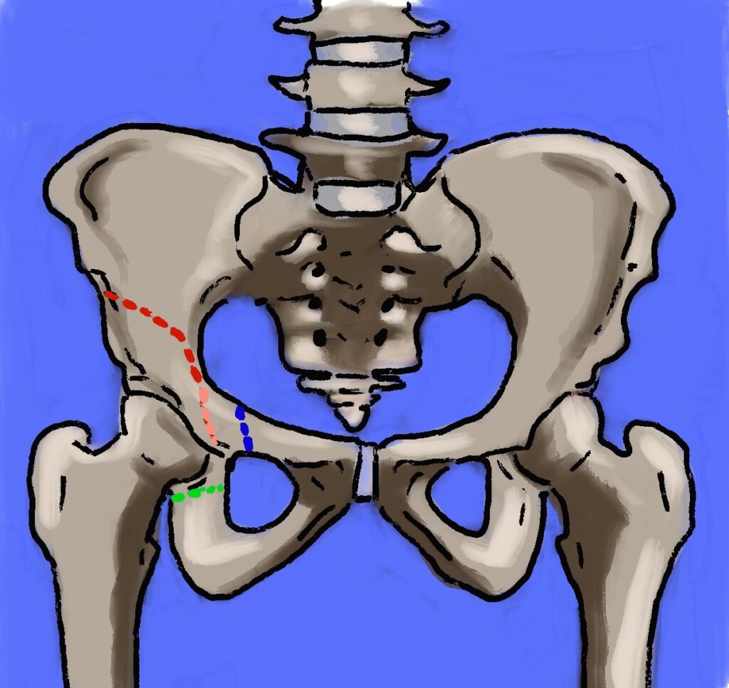
- Requires a concentric reduction for redirectional and reshaping osteotomies
- Centre edge angle under 20 degrees and an acetabular index above normal for age define dysplasia
- Pain from dysplasia in adolescents and young adults favours periacetabular osteotomy
Preoperative Assessment
- Concentric reduction on an abduction internal rotation view
- Range of motion and congruency, with arthrography in children
- CT for acetabular version and coverage
- MRI for labral and cartilage damage in adults
Classification
| Osteotomy | Type | Notes |
|---|---|---|
| Salter innominate | Redirectional | Single cut above the acetabulum hinging on the symphysis. Age 18 months to 6 years |
| Pemberton | Reshaping | Incomplete cut hinging on the triradiate cartilage. Reduces a capacious acetabulum |
| Dega | Reshaping | Incomplete cut with posterior hinge. Suits neuromuscular posterior deficiency |
| Triple (Steel, Tönnis) | Redirectional | Cuts through ilium, ischium and pubis. Older child |
| Bernese periacetabular (Ganz) | Redirectional | Closed triradiate cartilage. Posterior column preserved |
| Chiari | Salvage | Medial displacement osteotomy with capsular interposition for an incongruent hip |
| Shelf | Salvage | Bone graft extends the lateral acetabular roof |
Complications
- Sciatic and femoral nerve injury
- Over correction causing impingement
- Loss of correction and graft displacement
- Lateral femoral cutaneous nerve injury with anterior approaches
- Nonunion of pubic cuts
- Avascular necrosis of the femoral head with combined femoral osteotomy
- Heterotopic ossification
Reviewed by Professor Phong Tran, Head of Orthopaedic Surgery, Western Health. Last updated 10 October 2026.