
Reviewed by
Dr Andrew Mattin
MBBS | Accredited Orthopaedic Registrar
- 1:3000 newborns
- Equal in sex and race
- NF 1 – long arm of chromosome 17
- Gene product can be thought of as a tumour suppressor for cellular oncogene
- A neural crest disorder
- Cells migrate to parts of skin, brain, cord, nerves & adrenal glands
- Von Recklinghausen disease. (1882)
- Autosomal dominant disease. (Chromosome 17q11)
- Gene that codes for neurofibromin.
- NF 2 – Chromosome 22.(Schwannomin.)
- Less common (1 in 50,000) but also autosomal dominant.
- Mosaics of the 1 and 2 do not demonstrate orthopaedic manifestations
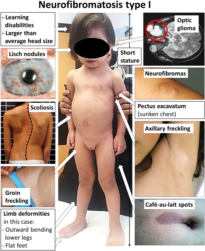
Classification
| Type | Features |
|---|---|
| NF 1 | 2 or more of the National Institute of Health criteria At least 6 café au lait spots (> 5mm in children, > 15 mm in adults) 2 neurofibromas or 1 plexiform neurofibroma Freckling of the inguinal or axillary region Optical glioma At least 2 Lisch nodules (iris condensations / hamartoma of the iris) A first degree relative with NF A distinctive osseous lesion (sphenoid dysplasia, thinning of long bone cortex with or without pseudarthrosis) |
| NF 2 | Bilateral acoustic neuromas (bilateral vestibular schwannomas). Paucity of peripheral findings |
- Diagnosis may be delayed due to variable age of presentation of signs
Clinical
- Café au lait spots – discrete tan spots
- May take up to 1 year to appear
- Smooth
- Café au lait macules.
- 99% have 6 or more that are >5cm in diameter by age of 1
- Skin fold freckling. Axillary, inguinal, upper eye lids. 90% at 7 years
- Neurofibroma
- Cutaneous – benign schwann cells with fibrous tissue
- Rapidly increasing numbers at puberty
- >80% at 20 years
- Plexiform neurofibromas
- present at birth, highly infiltrative, darkly pigmented, limb gigantism or facial disfigurement
- prone to haemorrhage, pain, disfiguration and malignant transformation.
- 10-24%.
- NF-1 lifetime risk of malignant transformation 5%.
- Axillary or inguinal freckling
- Optic pathway glioma
- Neurological delayed development
- HTN, CVA, congenital heart disease, vasculopathy.
Orthopaedic Manifestations
- Unusual scoliosis
- Scoliosis (Dystrophic, Non dystrophic)
- Nonunion of a long bone
- Overgrowth of a part
- Soft tissue tumours
General
- Osteopaenia complicates treatment
- Osteomalacia,
- Affects cellular growth of neural tissue
Spine
- Spinal deformity. 49% patients.
- intraspinal neurofibromatosis
- Dystrophic changes.
- Rib penciling,
- Dumbbell lesions
- Spindling of transverse process
- vertebral wedging
- foraminal enlargement
- widened interpedicular distance
- Dural ectasia
- Circumferential dilation of the thecal sac and erodes surrounding bony structures.
- Instability and deformity.
- T2 MRI
- Vertebral scalloping
- Anterior and posterior vertebral scalloping
- >3mm in thoracic spine, >4mm in lumbar spine.
- Posterior is most common
- 63% scalloped vertebrae have associated dural ectasia or intraspinal neurofibroma.
- Cervical spine
- 30% of NF-1 patients.
- Cervical kyphosis, rotatory subluxation, dysplastic changes.
- More common in dysplastic curves.
- Spondylolisthesis, spondyloptosis.
- Many are asymptomatic.
- Scoliosis
- Dystrophic
- Less common but more severe.
- Short sharp angulated scoliosis.
- Odd shaped vertebrae, codfish vertebrae
- Scalloped posterior body
- Elevated neural foramen
- Defective pedicles
- Dural abnormality on MRI
- Ribboned ribs
- Relentless progression
- Kyphosis common & can lead to neurologic dysfunction
- Non dystrophic
- Treat as idiopathic
- Progresses at same rate as adolescent idiopathic scoliosis and same clinic appearance.
- Earlier onset the worse prognosis.
- Must monitor due to progression.
- Management
- All should be assessed with an MRI and CT.
- Look for
- Intra/extra spinal neoplasm.
- Repeat if any rapid progression.
- Look for
- Treatment according to deformity
- Observation <20°
- Bracing 20-40°
- Fusing >40°
- Dystrophic scoliosis.
- High risk of progression and neurological compromise.
- Early and aggressive surgical intervention has been advocated to prevent dystrophic curve progression.
- Bracing has not been effective.
- 6 monthly monitoring <20°.
- 20-40° surgery.
- All should be assessed with an MRI and CT.
- Dystrophic
Nonunion
- May affect Tibia & fibula, radius, ulna, femur & clavicle
- Congenital pseudoarthrosis of the tibia (CPT)
- CPT 1 in 250,000 births.
- 5% NF-1 have CPT.
- Anterolateral bowing of tibia that presents in the first year of life.
- Cortical thickening with narrowed intramedullary canal.
- Spontaneous fracture followed by pseudarthrosis.
- Treatment
- Non operative.
- Knee-ankle-foot orthosis to prevent fracture in dysplastic bone.
- Operative
- Principles
- Variable union rates post operatively.
- High refracture rate.
- Common complication is non union, limb length discrepancy, refracture and valgus deformity.
- Options
- Bone grafting with IM fixation
- Telescoping nails
- EXFIX,
- Ring external fixator
- free vascularised fibular graft.
- Resection of pseudarthrosis site.
- Bone morphogenic proteins
- Bone grafting with IM fixation
- Principles
- Non operative.
Overgrowth
- Any child with local gigantism has NF until proven otherwise
- Overgrowth of some or all tissues in one region.
- Unilateral segmental hypertrophy or gigantism.
Other Lesions of Bone
- Benign scalloping of the cortex
- Cystic lesions
- Permeative bone destruction
Malignancy
- ~ 5%
- usually CNS
- neurofibrosarcoma can occur
Other
- Lisch nodules
- HTN secondary to renal artery stenosis
- 50% have cognitive impairment
- short big heads
Take-home Message
- NF-1 autosomal dominant. 1 in 3,000.
- Common orthopaedic manifestations which are challenging to manage.
- Multidisciplinary approach is warranted as multi-organ involvement
Reviewed by Professor Phong Tran, Head of Orthopaedic Surgery, Western Health. Last updated 10 October 2026.