
Classification
| Type | Causes |
|---|---|
| Primary (congenital) | Associated with vertebral anomalies (atlanto-occipital fusion, hypoplasia of the atlas, Klippel-Feil) Associated with skeletal dysplasias (achondroplasia, spondyloepiphyseal dysplasia, Morquio syndrome) |
| Secondary | Skull softening disorders (severe osteoporosis, osteomalacia, rickets, Paget’s, osteogenesis imperfecta, RA, neurofibromatosis) |
Pathology
Odontoid may impinge on cord
- medulla
- Cerebellum
- Vertebral arteries (VBI)
- Aqueduct of Sylvius (Hydrocephalus)
X-ray
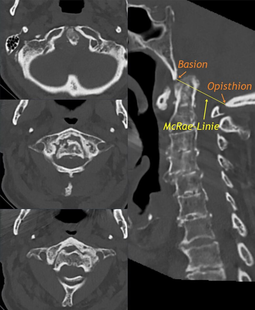
- McRaes line
- connects anterior + posterior margins of the foramen magnum
- (Basion + Opisthion)
- odontoid should not project above this line
- Chamberlains line
- From the posterior margin of the hard palate to the posterior margin of the foramen magnum
- Tip of the odontoid should not project more than 3mm above this line
- McGregors line
- From the posterior margin of the hard palate to the most caudal part of the occiput
- tip of the odontoid should not project > 4.5 mm above this line
Treatment
- Halo
- Preop Halo traction to reduce subluxations & pull the odontoid out of the foramen magnum
- Occipito-cervical fusion
- Technique
- midline incision – from extension occip protuberance to ~ C4 level
- subperiosteal dissection to expose occiput & cervical laminae
- wires passed
- under C1 laminae
- through drill hole in C2 SP
- through hole in outer table at external occipital protuberance
- decorticate + make trough in occiput for graft
- BG from posterior iliac crest harvested, laid, wires passed through drill holes in graft & then tied over
- cervical orthosis for 3/12
Reviewed by Professor Phong Tran, Head of Orthopaedic Surgery, Western Health. Last updated 10 October 2026.