
Question
A 12 year old boy has recurrent ankle sprains and a painful, stiff flat foot. The subtalar joint has very little motion.
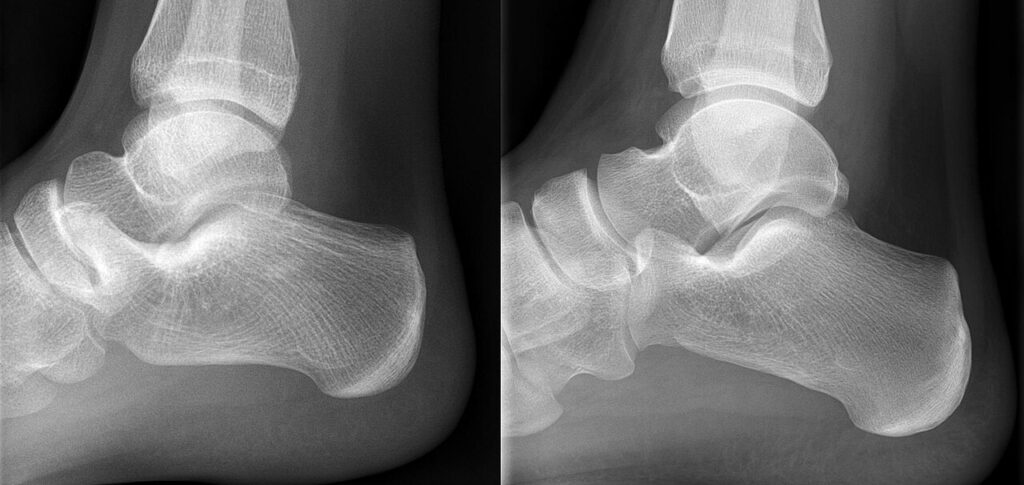
- What is the likely diagnosis and how would you confirm it?
- What are the radiographic signs?
- Outline management.
Answer
Diagnosis
- Tarsal coalition causing a rigid flat foot, most often calcaneonavicular or talocalcaneal (middle facet)
- Failure of segmentation of the mesenchyme, autosomal dominant, bilateral in about half
- Calcaneonavicular coalitions present around 8 to 12 years and talocalcaneal around 12 to 16 years as they ossify
- Heel valgus does not correct on tiptoe and the subtalar joint is stiff, with peroneal spasm
Radiographic signs
- Anteater nose sign on the lateral view for calcaneonavicular coalition, best seen on the 45 degree oblique view
- C sign and talar beaking on the lateral view for talocalcaneal coalition
- Harris axial view for the middle facet
- CT confirms and maps the coalition, and MRI shows fibrous and cartilaginous coalitions
Management
- Activity modification, orthoses and a short leg walking cast for 4 to 6 weeks
- Resection of calcaneonavicular coalition with interposition of extensor digitorum brevis or fat
- Resection of talocalcaneal coalition involving less than about 50% of the posterior facet, with fat interposition
- Triple arthrodesis for failed resection, large coalitions or established arthritis
Related pages
Author Contributions
Orthofracs team
Reviewed by Professor Phong Tran, Head of Orthopaedic Surgery, Western Health. Last updated 10 October 2026.