
Indications
- True internervous approach to the anterior hip and ilium
- Open reduction of developmental dysplasia of the hip
- Drainage of septic arthritis
- Femoral head fractures, especially anterior Pipkin fragments
- Salter osteotomy, periacetabular osteotomy and iliac crest graft harvest
- Hemiarthroplasty and total hip replacement, with DAA using the distal interval
Position
- Supine with a small bolster under the ipsilateral buttock
Landmarks and Incision
- Landmarks are the iliac crest and ASIS
- Incision along the anterior half of the crest to the ASIS, then distally towards the lateral patella
Internervous Plane
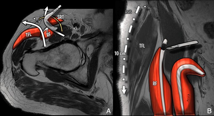
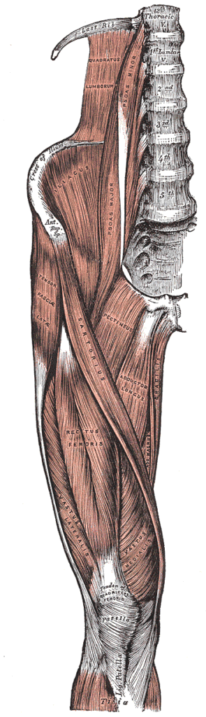
- Superficial plane between sartorius (femoral nerve) and TFL (superior gluteal nerve)
- Deep plane between rectus femoris (femoral nerve) and gluteus medius (superior gluteal nerve)
Superficial Dissection
- LFCN pierces the fascia near the ASIS and runs over sartorius, so incise fascia on the tensor side
- Develop the sartorius and TFL interval
- Proximally elevate gluteus medius and TFL subperiosteally from the outer table
- Ligate the ascending branch of the lateral femoral circumflex artery
Deep Dissection
- Detach rectus femoris direct head from the AIIS and reflected head from the superior rim
- Retract rectus and iliopsoas medially and gluteus medius laterally
- T-shaped capsulotomy
- Anterior dislocation with external rotation and adduction
Direct anterior approach
- Distal Hueter interval through a shorter incision, with no muscle detached from the pelvis
- Incision distal and lateral to the ASIS over the TFL belly
- TFL retracted laterally within its sheath, keeping the LFCN medial
- Neck osteotomy in situ, with fluoroscopy for cup position and leg length
- Femoral exposure needs extension, adduction, external rotation and posterosuperior capsular release
- Muscle sparing with a low dislocation rate
- LFCN neurapraxia, femoral fracture or perforation, and wound problems in obese groin folds
Structures at Risk
- LFCN, causing meralgia paraesthetica
- Femoral nerve, medial to the approach
- Ascending branch of the lateral femoral circumflex artery
Extension
- Proximally along the crest into the iliofemoral approach to both iliac tables
- Distally into the anterolateral approach to the femoral shaft
Reviewed by Professor Phong Tran, Head of Orthopaedic Surgery, Western Health. Last updated 10 October 2026.