
Question
A 70 year old man has thigh pain 15 years after total hip arthroplasty. Infection markers are normal.
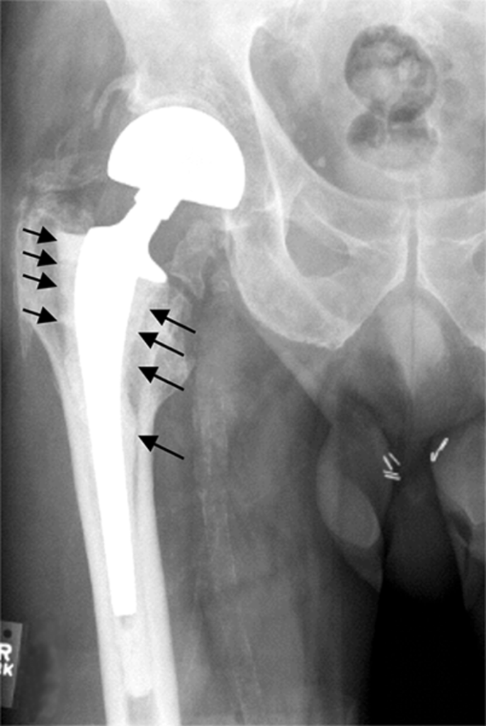
- Describe the radiograph.
- Explain the biology of osteolysis.
- How do you assess and manage this patient?
Answer
Radiograph
- Radiolucent lines and endosteal scalloping around the femoral stem (arrows)
- Femoral component loosening with osteolysis
- Gruen zones describe the femoral location, DeLee and Charnley the acetabular
Biology
- Polyethylene wear particles 0.1 to 1 micron
- Macrophage phagocytosis with release of TNF alpha, IL1 and IL6
- RANKL mediated osteoclast activation
- Effective joint space allows particles to spread along interfaces
- Highly cross linked polyethylene has reduced osteolysis
Assessment
- Exclude infection with aspiration
- Serial radiographs for progression
- CT to measure lysis and bone stock
- Paprosky classification of femoral and acetabular bone loss
Management
- Asymptomatic stable implants with small lesions, observe
- Well fixed cup with lysis, liner exchange and grafting
- Loose stem, revision with distal fixation (modular tapered or extensively coated stem)
- Impaction grafting is an option
- Extended trochanteric osteotomy for well fixed cement or stems
Related pages
Author Contributions
Orthofracs team
Reviewed by Professor Phong Tran, Head of Orthopaedic Surgery, Western Health. Last updated 10 October 2026.