
Question
A 6 year old girl falls from a trampoline. Her hand is pink and warm with a radial pulse, and she cannot make an OK sign.
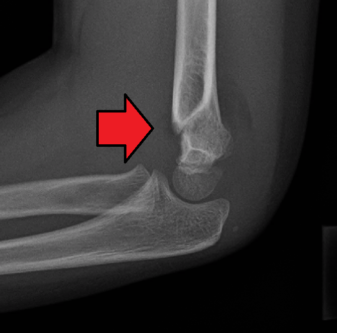
- Describe the radiograph and classify the injury.
- Which nerve is injured and how do you manage it?
- Describe closed reduction and pinning.
Answer
Radiograph
- Extension type supracondylar fracture of the humerus
- Anterior humeral line misses the middle third of the capitellum
- Posterior fat pad visible
Gartland classification
| Type | Description |
|---|---|
| I | Undisplaced |
| II | Displaced, posterior cortex intact |
| III | Completely displaced |
| IV | Unstable in flexion and extension (Leitch) |
Nerve injury
- Anterior interosseous nerve, the commonest nerve injury in extension types
- Cannot flex the IP joint of the thumb or DIP joint of the index finger
- Posteromedial displacement injures the radial nerve, posterolateral the median nerve and brachial artery
- Ulnar nerve injury is often iatrogenic from a medial pin
- Most recover over 2 to 3 months, document before surgery and observe
Closed reduction and pinning
- Traction, correct varus or valgus and rotation, then flex with pressure on the olecranon
- Pronate for posteromedial displacement
- Two or three lateral divergent pins, cross the fracture and engage both columns
- Medial pin only through a small incision with the elbow extended
- Above elbow backslab in under 90 degrees flexion
- Remove pins at 3 to 4 weeks
Related pages
Author Contributions
Orthofracs team
Reviewed by Professor Phong Tran, Head of Orthopaedic Surgery, Western Health. Last updated 10 October 2026.