
Question
A 7 year old girl has a painless clunk in her knee with loss of full extension.
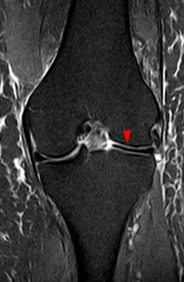
- What does the MRI show?
- Describe the classification.
- Outline management.
Answer
MRI
- Thickened lateral meniscus covering most of the lateral tibial plateau
- Discoid lateral meniscus
- Continuity of the meniscus across three or more consecutive 5 mm sagittal slices (bow tie sign)
- Bilateral in up to 20%
Watanabe classification
| Type | Description |
|---|---|
| I | Complete, covers the whole plateau, stable |
| II | Incomplete, stable |
| III | Wrisberg variant, no posterior tibial attachment, unstable |
- Ahn classification describes the direction of meniscal shift on MRI
Management
- Asymptomatic discoid menisci are observed
- Arthroscopic saucerisation for symptomatic tears, leaving a 6 to 8 mm rim
- Repair the peripheral rim if unstable
- Avoid total meniscectomy, which leads to early lateral compartment arthritis
- Osteochondritis dissecans of the lateral femoral condyle can coexist
Related pages
Author Contributions
Orthofracs team
Reviewed by Professor Phong Tran, Head of Orthopaedic Surgery, Western Health. Last updated 10 October 2026.