
Question
A 20 year old footballer had an ankle injury treated surgically. These are his postoperative radiographs.
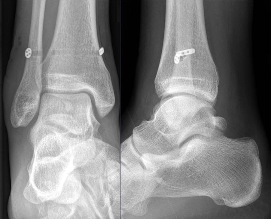
- What injury has been treated and how is it diagnosed?
- Compare screw and suture button fixation.
- What is a Maisonneuve injury?
Answer
Injury and diagnosis
- Syndesmosis disruption treated with a suture button (TightRope)
- High ankle sprain, external rotation mechanism
- Squeeze test and external rotation test
- Radiographic tibiofibular clear space over 6 mm, reduced tibiofibular overlap, medial clear space widening
- Weight bearing or stress views, MRI or bilateral weight bearing CT if uncertain
- Arthroscopy is the most accurate test of instability
Screw versus suture button
| Feature | Screw | Suture button |
|---|---|---|
| Malreduction | More common | Less common, allows self correction |
| Removal | Often removed or breaks | Rarely removed |
| Function | Similar or slightly worse | Similar or slightly better early |
- Malreduction is the main predictor of poor outcome
- Direct visualisation of the incisura or intraoperative CT improves reduction
- Clamp in neutral rotation along the transmalleolar axis
Maisonneuve injury
- Proximal fibular fracture with syndesmosis and deltoid ligament or medial malleolar injury
- Always examine the proximal fibula
- Peroneal nerve at risk
- Treated by syndesmotic fixation without fibular fixation
Related pages
Author Contributions
Orthofracs team
Reviewed by Professor Phong Tran, Head of Orthopaedic Surgery, Western Health. Last updated 10 October 2026.