
Question
A 45 year old man rolls his ankle on a step. The ankle is deformed with tented skin medially.
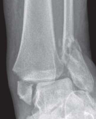
- Describe the radiograph and your immediate management.
- Classify the injury.
- What are the principles of fixation?
Answer
Radiograph and immediate care
- Bimalleolar fracture dislocation of the ankle
- Talar shift
- Urgent reduction in the emergency department to protect the skin
- Backslab, elevation and post reduction films
- Check neurovascular status before and after
Classification
| System | Category |
|---|---|
| Weber | Fibula at the level of the syndesmosis is Weber B |
| Lauge-Hansen | Supination external rotation IV is the commonest pattern |
| AO | 44 B |
Fixation principles
- Swelling may delay surgery, fix when skin wrinkles
- Restore fibular length and rotation
- Lag screw and neutralisation plate or posterior antiglide plate for the fibula
- Medial malleolus with two partially threaded screws or tension band
- Test the syndesmosis after fixation (Cotton or stress test)
- Fix posterior malleolar fragments that involve the incisura or cause instability
- Diabetic patients need more rigid fixation and longer protection
Evidence
- AIM trial showed close contact casting was equivalent to surgery in patients over 60 with unstable fractures
Related pages
Author Contributions
Orthofracs team
Reviewed by Professor Phong Tran, Head of Orthopaedic Surgery, Western Health. Last updated 10 October 2026.