
Question
A 38 year old motorcyclist has a painful swollen knee after a crash. Plain radiographs were reported as showing only an effusion.
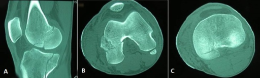
- Describe the CT and name the fracture.
- Why is it easily missed and how is it classified?
- How would you fix it?
Answer
CT
- Coronal plane fracture of the posterior femoral condyle
- Hoffa fracture (AO 33 B3)
- Lateral condyle more common
Missed diagnosis
- Fracture plane is coronal, so the AP radiograph often looks normal
- A true lateral may show a double contour
- CT for all distal femoral fractures, as Hoffa fragments occur in up to 38% of intercondylar fractures
Letenneur classification
| Type | Description |
|---|---|
| I | Whole posterior condyle, line parallel to the posterior femoral cortex |
| II | Basal fragment, subtypes a to c by size |
| III | Oblique fracture |
Fixation
- Unstable and displaced, so non-operative care fails
- Anatomical reduction through a lateral parapatellar or posterolateral approach
- Anterior to posterior lag screws perpendicular to the fracture, countersunk
- Posterior to anterior screws are stronger
- Buttress plate for basal or comminuted fragments
- Early motion
Related pages
Author Contributions
Orthofracs team
Reviewed by Professor Phong Tran, Head of Orthopaedic Surgery, Western Health. Last updated 10 October 2026.