
Question
A 24 year old female military recruit has activity related groin pain that has worsened over three weeks of training.
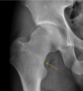
- Describe the radiograph and the next investigation.
- How do you classify these fractures?
- Outline management and risk factors to address.
Answer
Radiograph
- Sclerosis on the inferomedial (compression side) femoral neck
- Femoral neck stress fracture
- Radiographs are often normal early
- MRI is the investigation of choice
Classification
| Type | Management |
|---|---|
| Compression side, under 50% of neck width | Protected weight bearing |
| Compression side, over 50% or progression | Fixation |
| Tension side (superolateral) | Fixation, high risk of displacement |
| Displaced | Urgent reduction and fixation |
Management
- Non weight bearing with crutches for compression fractures
- Repeat imaging to confirm healing
- Cannulated screws for tension side fractures
- Displaced fractures in young adults need urgent anatomical reduction and fixation
- AVN and nonunion are common after displacement
Risk factors
- Relative energy deficiency in sport, the female athlete triad
- Vitamin D deficiency
- Sudden training increase
- Coxa vara
Related pages
Author Contributions
Orthofracs team
Reviewed by Professor Phong Tran, Head of Orthopaedic Surgery, Western Health. Last updated 10 October 2026.