
Question
A 40 year old man on long term corticosteroids for asthma has 3 months of groin pain. Radiographs are normal.
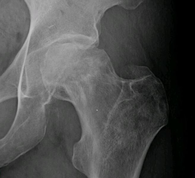
- What is the likely diagnosis and what are the risk factors?
- How would you investigate and stage it?
- Outline management by stage.
Answer
Diagnosis and risk factors
- Osteonecrosis of the femoral head (AVN)
- Risk factors include corticosteroids, alcohol, trauma, sickle cell disease, Gaucher disease, caisson disease, SLE and HIV
- Bilateral in up to 50 to 80% of non-traumatic cases
Investigation and staging
- MRI of both hips, which shows the double line sign and is sensitive before radiographic change
- Ficat and Arlet or Steinberg staging
- Size of the necrotic lesion (Kerboul angle) and location predict collapse
- Look for a crescent sign of subchondral fracture on frog lateral radiographs
Management by stage
- Stop or reduce risk factors where possible
- Pre-collapse small lesions, observation or core decompression, with or without bone graft or cell therapy
- Pre-collapse larger lesions, core decompression with structural or vascularised fibular graft in selected young patients
- Rotational or angular osteotomy in selected patients with limited lesions
- Post-collapse and arthritic hips, total hip arthroplasty, with outcomes now similar to osteoarthritis
Related pages
Author Contributions
Orthofracs team
Reviewed by Professor Phong Tran, Head of Orthopaedic Surgery, Western Health. Last updated 10 October 2026.